
- Title
-
Developmental roles of D-bifunctional protein-A zebrafish model of peroxisome dysfunction
- Authors
- Kim, Y. I., Bhandari, S., Lee, J. N., Yoo, K. W., Kim, S. J., Oh, G. S., Kim, H. J., Cho, M., Kwak, J. Y., So, H. S., Park, R., Choe, S. K.
- Source
- Full text @ Mol. Cells
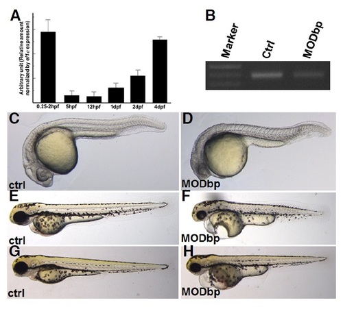
dbp expression is essential for zebrafish embryogenesis. (A) dbp expression during development. Quantitative RT-PCR was used to assay dbp expression during development. The highest expression of dbp is at 0-2 hpf (maternally deposited) after which it is significantly decreased but increased gradually at 1dpf onwards. The graph shows relative amount of dbp mRNA normalized by ef1± expression. (B) dbp splicing-blocking morpholino (MO) efficiently reduced synthesis of mature mRNA. Total RNA extracted from control and MO-injected embryos at 1 dpf were used for cDNA synthesis followed by RT-PCR. A ~ 390 bp band in the control lane was significantly reduced in the morpholino-injected lane (MODbp), indicating high efficiency of MO. (C-H) dbp knockdown generates morphologically distinct phenotypes. MO-injected embryos displayed phenotypes, includingsmall head, pericardial edema, and voluminous yolk compared to control embryos [compare (C, E, and G) with (D, F, and H)]. As embryogenesis continued, phenotypes became more severe [(C) and (D) (1dpf); (E) and (F) (2 dpf); (G) and (H) (3 dpf)]. Embryos are shown in lateral view with anterior to the left. PHENOTYPE:
|
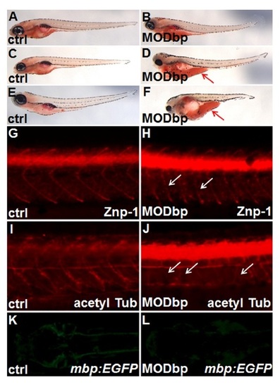
dbp knockdown affects yolk consumption and neuronal development. (A-F) Embryos with dbp knockdown showed accumulated neutral lipids in the yolk. Oil Red-O staining revealed a significant delay or defect in yolk lipid consumption in dbp knockdown embryos on or after 3 dpf, (A) and (B) (3 dpf); (C) and (D) (4 dpf); (E) and (F) (5 dpf). As compared to control embryo, greater quantities of yolk lipids (visualized in red) were retained in a dbp knockdown embryo. Red arrows in (D) and (F) indicate accumulated lipids in the yolk. (G-L) dbp knockdown impaired neuronal development. Immunostaining using anti-Znp-1 antibody (G, H) or anti-acetylated tubulin (I, J) revealed defective neuronal development, such as discontinued axonal projections [indicated by white arrows in (H)] or suppression of differentiating motor axons [indicated by white arrows in (I)] upon dbp knockdown. (K, L) GFP expression driven by the myelin basic protein (mbp) promoter (indicative of myelinating cells) in an mbp:EGFP transgenic line was significantly reduced following dbp knockdown. Embryos are shown in lateral view with anterior to the left, except in (K) and (L) where embryos are in dorsal view. Partial trunk regions of embryos are shown in (G-J). EXPRESSION / LABELING:
PHENOTYPE:
|
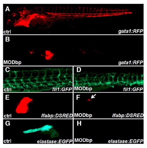
dbp knockdown affects development of blood, blood vessels and digestive organs. Embryos carrying different transgenes were used to determine tissue-specific effects upon dbp knockdown. (A-D) dbp knockdown affects red blood cell development and blood vessel patterning. Upon dbp knockdown, blood cells were severely reduced [shown by gata1:RFP transgenic line, (A) and (B)] and blood vessels were formed abnormally [shown by fli1:EGFP, (C) and (D)]. (E-H) dbp knockdown on doubly transgenic fish line (lfabp: DSRED in liver and elastase:GFP in pancreas) shows significantly reduced liver size (F) and absence of pancreas (H). Representative embryos at 3 dpf are shown in lateral view with anterior to the left, except (G) and (H) where presented in lateral view with anterior to the right. PHENOTYPE:
|

Knockdown of dbp affects expression of genes involved in peroxisome functions and mitochondrial biogenesis. (A-F) Quantitative RT-PCR was used to assay expression of genes involved in ether phospholipid synthesis: gnpat1 (A) and agps(B), a gene in peroxisomal protein import: pex5 (C),and genes in mitochondrial biogenesis: pgc1a (D), pparab (E) and esrra (F). dbp knockdown altered the expression of genes in involved in ether phospholipid synthesis starting at 1 dpf (A-B) and that of genesin peroxisomal protein transport or mitochondrial biogenesis at 2 dpf. * indicates statistical significance (p < 0.05) between control and dbp MO samples. (G) DBP is functionallyconserved between mice and zebrafish. Rescue experiment was performed by co-injecting murine Dbp mRNA together with dbp MO. Embryos belonging to each category of phenotypic severity shown in the right were counted from three independent experiments and presented as percentage of total injected embryos. EXPRESSION / LABELING:
PHENOTYPE:
|