
- Title
-
Use of antagonists and morpholinos in loss-of-function analyses: estrogen receptor ESR2a mediates the effects of 17alpha-ethinylestradiol on primordial germ cell distribution in zebrafish
- Authors
- Hu, J., Sun, S., Guo, M., Song, H.
- Source
- Full text @ Reprod. Biol. Endocrinol.
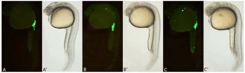
Fluorescence images of zebrafish PGCs after exposure to various concentrations of EE2. A: In all treatment groups, normal PGCs were observed in the anterior region of the yolk extension at 24 hpf. B: In the 1 ng/L, 10 ng/L, and 100 ng/L EE2 exposure groups, ectopic PGCs were primarily observed along the branchial arch, similarly to the spadetail mutant. C: In the 500 ng/L, 1 μg/L and 2 μg/L EE2 exposure groups, many ectopic PGCs were observed along the branchial arch, on the back, in the abdomen, and along the trunk. A′, B′, and C′: Bright-field views show the morphology of the embryos. |
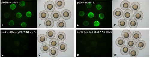
Effects of MO on GFP expression in 7 hpf embryos injected with recombinant plasmids. A, B: A mosaic pattern of GFP expression was detected throughout the embryos. C, D: GFP expression was undetectable after co-injection with MO and recombinant plasmids. A′, B′, C′, D′: Bright-field views show the morphology of the embryos. |