
- Title
-
transparent, a gene affecting stripe formation in Zebrafish, encodes the mitochondrial protein Mpv17 that is required for iridophore survival
- Authors
- Krauss, J., Astrinidis, P., Frohnhöfer, H.G., Walderich, B., and Nüsslein-Volhard, C.
- Source
- Full text @ Biol. Open
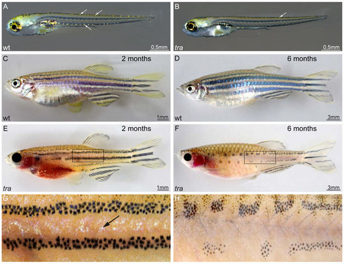
transparent mutants show a reduction in iridophore pigmentation. (A,C,D) Wild type and (B,E-H) tra mutants. At 5 dpf, mutant larvae show strong reduction in iridophores in dorsal and ventral positions (B) as compared to wild type (A). Arrows in panels A and B highlight the appearance and position of iridophores in larvae. No other defects are apparent. Two months old wild type animals (C) present a fully developed pigment pattern, while tra individuals (E) developed only few iridophores, visible between the dark stripes. (G) Close-up of the region boxed in panel E. At that age, melanophores are reduced in number in the mutants, however, the typical four stripes developed. While the mutant fish grow, the melanophore stripes break up into spots (F, close up in H), compare six months old tra mutant (F) to wild type (D) of the same age. The abdominal cavity, typically covered by a thick sheet of iridophores in wild type, lacks this cell type in tra mutants. Iridophores of the eyes are strongly reduced throughout life in the mutants. Pigment patterning of fins and scales appear normal. Scale bars: 0.5 mm (A,B); 1 mm (C,E), 3 mm (D,F). |
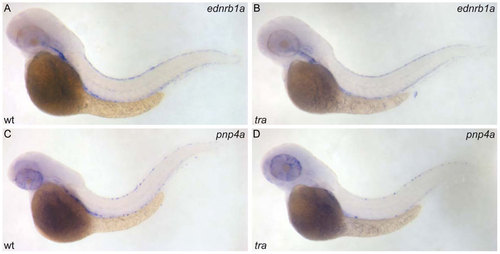
transparent affects pigment synthesis of iridophores, but not their specification and initial differentiation. ednrb1a RNA hybridization of wild type (A) and tra mutant larva (B), pnp4a RNA hybridization of wild type (C) and tra mutant (D); 48 hpf. EXPRESSION / LABELING:
|
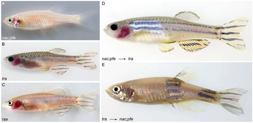
transparent is required cell-autonomously in iridophores. (A-C) Mutants used in transplantation experiments: (A) nac;pfe, (B) tra, (C) rse. (D) nac;pfe mutant which received tra mutant xanthophores and melanophores, showing that both cell types contribute normally to stripes. (E) tra mutant which received nac;pfe mutant iridophores, showing that the supply with iridophores is sufficient to normalize the pigment pattern. |
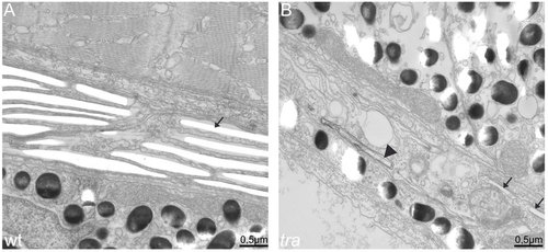
transparent mutant iridophores display characteristics of apoptotic cells. EM sections of wild type (A) and transparent mutant iridophores (B) in the eye of 5 dpf larvae. (A) Wild type iridophores develop stacks of iridosomes nearly completely filling the cells. Iridosomes are not contrasted and appear empty as the guanine crystals are lost during the sectioning process (arrow). (B) Iridosomes are also visible in tra mutants. In the cell shown, only two iridosomes seem to contain guanine crystals (arrows), whereas others contain contrastable material (arrowhead), indicating that guanine deposition is not completed. Importantly and in contrast to wild type, tra mutant iridophores contain many vesicles, a sign for apoptosis. Scale bars: 0.5 mm. PHENOTYPE:
|