
- Title
-
The DMAP interaction domain of UDP-GlcNAc:lysosomal enzyme N-acetylglucosamine-1-phosphotransferase is a substrate recognition module
- Authors
- Qian, Y., Flanagan-Steet, H., van Meel, E., Steet, R., and Kornfeld, S.A.
- Source
- Full text @ Proc. Natl. Acad. Sci. USA
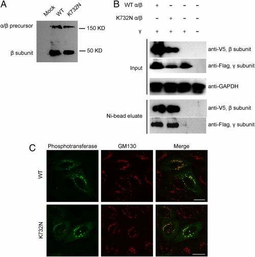
The K732N mutant exhibits normal expression, interaction with the γ subunit, processing, and localization. (A) HEK293 cells were transfected with plasmids encoding WT or K732N mutant α/β cDNAs with a C-terminal His/V5 tag. After 48 h, cell extracts were prepared and subjected to SDS/PAGE and immunoblotting with anti-V5 antibody to detect the α/β precursor and the cleaved β subunit. (B) Lysates of HEK293 cells expressing WT or K732N mutant α/β subunits with His/V5 tags plus γ subunits with a Flag tag or γ subunit alone were incubated with Ni-NTA agarose to bind the β subunit. Bound proteins were eluted and subjected to SDS/PAGE and immunoblotting with anti-V5 to detect the β subunit and anti-Flag to detect the γ subunit. (Upper) The expression of proteins in the original lysates (Input), with GAPDH serving as a loading control. (Lower) The proteins bound and eluted from the Ni-NTA resin (Ni-bead eluate). Nontransfected cells served as an additional control. (C) HeLa cells were transfected with WT (Upper) or K732N (Lower) α/β cDNA in pcDNA6 and fixed with 4% paraformaldehyde/PBS 16 h after transfection. The cells were stained for immunofluorescence microscopy with antibodies to the alpha subunit of phosphotransferase (green) and the cis-Golgi marker GM130 (red). (Scale bars, 20 μm.) |
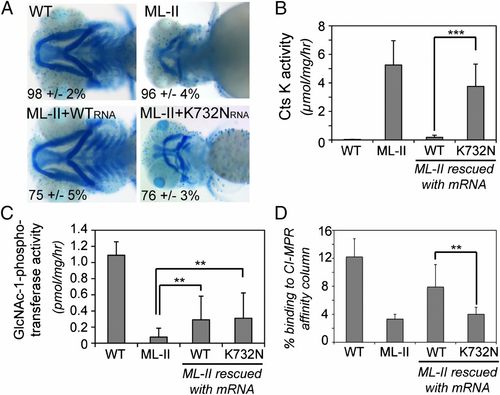
The K732N mutant fails to rescue MLII zebrafish. (A) Embryos were stained at 4 dpf with Alcian blue to reveal cartilage; representative images are shown. The percentage of embryos with a staining pattern consistent with the images shown is presented. Results represent an average of 5 individual experiments, with 30–40 embryos scored per experiment. (B) Cathepsin K activity was measured in embryo lysates, using a fluorogenic peptide substrate. Average activity from 8 to 9 independent experiments is shown. Error bars represent SD for all of the graphs. ***P < 0.01. (C) GlcNAc-1-phosphotransferase activity toward αMM was determined in embryo lysates, using the UDP-[3H]GlcNAc substrate. Results represent the average of 3 independent runs. **P < 0.05. (D) Bars represent the percentage of β-galactosidase activity that is Man-6-P modified in embryo lysates that were fractionated using a CI-MPR affinity column. Averages from 4–5 individual experiments are shown. **P < 0.05. PHENOTYPE:
|