
- Title
-
Identification and characterization of the zebrafish pharyngeal arch-specific enhancer for the basic helix-loop-helix transcription factor Hand2
- Authors
- Iklé, J.M., Artinger, K.B., and Clouthier, D.E.
- Source
- Full text @ Dev. Biol.
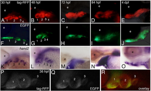
Both zebrafish and mouse enhancers drive transgene expression in a hand2-specific pattern within the pharyngeal arches. Dorsolateral views are shown at 30 and 48 hpf, and lateral views at 72 and 84 hpf and 4 dpf. A–E. Tg(hand2:tag-RFP) embryos were examined for RFP fluorescence between 30 h post fertilization (hpf) and 4 day post-fertilization (dpf). RFP was first detected at 30 hpf in the ventral aspect of pharyngeal arches 1–3 (A). At 48 hpf, RFP was present in the ventral aspects of arches 1–4 (B). At 72 hpf, RFP also appeared ventrally located (C). By 84 hpf (D) and 4 dpf (E), RFP was present in the developing cartilages while also appearing to expand dorsally. F-J. Tg(mHand2:EGFP) embryos were examined at the same stages as the Tg(hand2:tag-RFP) embryos. EGFP activity was observed in a hand2 specific expression pattern in the ventral aspects of arches 1–3 beginning around 30 hpf (F) and persisting in arches 1–4 through 48 hpf (G). EGFP activity continued as chondrogenesis began around 72–84 hpf (H, I) and was still present at 4 dpf (J), though EGFP activity expanded dorsally at both 84 hpf and 4 dpf. K–O. Whole mount in situ hybridization analysis of hand2 expression. Endogenous hand2 expression was present in arches 1–4 at 30 hpf (K) and 48 hpf (L). While endogenous hand2 expression remained at 72 and 84 hpf (M, N), expression began to be restricted by 4 dpf (O). P–R. Compressed confocal z-stacks of Tg(hand2:tag-RFP), Tg(mHand2:EGFP) embryos at 36 hpf in a lateral view, illustrating localization of tag-RFP (P), EGFP (Q) and co-localization of both reporters (R) within the ventral pharyngeal arches. |
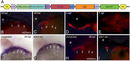
The minimal hand2 arch-specific enhancer is regulated by endothelin signaling. A. A 280 bp region of sequence contained within the larger zhand2 enhancer shown in Fig. 1C was cloned into p5E-FAbas and recombined with a middle entry clone containing a mCherry fluorescent reporter into the Tol2 destination vector pDestTol2CG2, resulting in hand2-directed mCherrry expression. Plasmid is not drawn to scale. B–E. In Tg(shand2:mCherry) embryos, mCherry fluorescence was observed in the Hand2 domain beginning around 30 hpf (B) and continuing through arch development (C, D). mCherry fluorescence can still be detected in the ventral cartilages as late as 7 dpf (E). F, G. Injecting 5 ng of an edn1 morpholino led to decreased hand2 expression at 48 hpf (G) compared to wild-type expression (F). H, I. mCherry activity was similarly decreased in Tg(shand2:mCherry) embryos following injection of the edn1 morpholino (I) compared to uninjected Tg(shand2:mCherry) embryos (H). |
Reprinted from Developmental Biology, 368(1), Iklé, J.M., Artinger, K.B., and Clouthier, D.E., Identification and characterization of the zebrafish pharyngeal arch-specific enhancer for the basic helix-loop-helix transcription factor Hand2, 118-126, Copyright (2012) with permission from Elsevier. Full text @ Dev. Biol.