
- Title
-
Visualization of gli activity in craniofacial tissues of hedgehog-pathway reporter transgenic zebrafish
- Authors
- Schwend, T., Loucks, E.J., and Ahlgren, S.C.
- Source
- Full text @ PLoS One
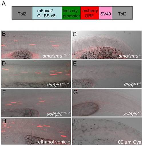
Gli-dependent reporter is capable of detecting modulations to the Hh-signaling pathway when expressed transiently in fish. (A) Schematic of the Gli-d mcherry reporter construct designed in this study. The construct consists of a 0.7 kb fragment encoding eight repeating Gli-CBS from the mouse FoxA2 floor plate enhancer and a minimal lens crystallin promoter (light blue and green respectively) inserted upstream of the mCherry ORF (red). The SV40 polyA signal (pink), which contains a transcriptional termination element, is directly downstream of the mCherry ORF and the entire element is flanked by Tol2 transposable elements (gray). (B–I) Lateral views, anterior to the left, of confocal stack projections of fixed trunk tissue. (B–G) Homozygous genetic mutants (-/-) for smu/smo (B,C), dtr/gli1 (D,E), yot/gli2 (F,G) were sorted from heterozygotes (+/-) and homozygous wild type (+/+) siblings based on evident characteristic Hh-signaling mutant phenotypes. Genetic mutants (-/-) for Hh-signaling pathway components failed to express mCherry protein (C,E,G), while strong mCherry expression was evident in trunk tissue of (+/+,+/-) siblings (B,D,F). (H,I) Hh-pathway attenuation with 100 µm Cya fully reduced mCherry protein expression in construct-expressing embryos (I) compared to vehicle-treated controls (H). PHENOTYPE:
|

Early expression of the mCherry reporter in transgenic embryos. RT-PCR analysis of mCherry from offspring of female or male Tg(Gli-d:mCherry) and wild type adults at stages ranging from 0.25 hpf up to 8 hpf. Expression of gapdh served as a cDNA loading control. |
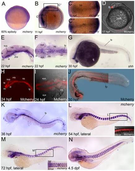
mCherry reporter gene and protein expression reflects known Hh-expressing domains in Tg(Gli-d:mCherry). Lateral views (A,B,D,E,G,H,J,K,L,L2,M,N) or dorsal views (C′,C",F,I,M′), anterior to the left, of Tg(Gli-d:mCherry) fish labeled with mCherry (A–F,K,L,M,N) or shh (G) riboprobes or mCherry protein which was visualized by confocal microscopy (H–J,L′). (A) mCherry is broadly expressed in blastomeres at 60% epiboly. (B–C") By 11 hpf, mCherry was localized to the neural plate (np) and the midline notochord (no) tissues. (D) At 17 hpf, mCherry protein was apparent in forebrain (fb) and floor plate (fp). (E–G) At 22–24 hpf, mCherry gene (E,F) and protein (H–J) are expressed in the fb, midbrain and fp, similar to shh gene expression (G) at a comparable stage. (K–N) At later stages from 2 to 4 dpf, mCherry is shown in whole-embryo lateral views and detectable in brain, fp, myotomal adaxial cells and PA. (L′) Confocal projections of Mcherry protein corresponding to boxed region in panel L revealed expression in the adaxial cells. (M′) Dorsal view of boxed region in panel M showed mCherry expression in adaxial cells and in somitic tissue neighboring the notochord in an orderly, segmental fashion. Abbreviations: di, diencephalon; fb, forebrain; fmb, forebrain-midbrain boundary; fp, floor plate; hb, hindbrain; mb, midbrain; np, neural plate; no, notochord; os, optic stalk; ov, otic vesicle; PA, pharyngeal arches; pcp, prechordal plate; te, telencephalon. |
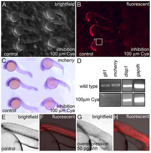
Reporter expression in Tg(Gli-d:mCherry) fish is sensitive to the Hh-signaling pathway. (A–C) Lateral views, anterior to the left, of 30 hpf ethanol vehicle treated controls (left side of image) and Cya treated (right side) Tg(Gli-d:mCherry) siblings. (A,B) Live embryos were visualized using brightfield (A) and fluorescence (B) microscopy. 100 &mu:m Cya treated embryos showed a significant reduction in fluorescent mCherry protein when compared to their ethanol vehicle treated siblings. (C) Fixed embryos, stained with mCherry riboprobe revealed that 100 μm Cya treated embryos displayed significantly reduced mCherry RNA transcripts compared to their vehicle-treated siblings. (D) RT-PCR showing reduced gli1, and mCherry expression in 30 hpf embryos treated with 100 μm Cya compared to their nontreated (NT) siblings. Two loading controls for disp1 and gapdh were included. (E–H) Lateral views of 30 hpf noninjected controls (E,F) or Tg(Gli-d:mcherry) siblings injected with 50 pg shh at the one cell stage (G,H). Siblings injected with shh mRNA showed expanded reporter expression throughout the trunk when compared to noninjected controls. EXPRESSION / LABELING:
|
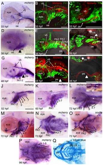
Reporter expression in Tg(Gli-d:mCherry) craniofacial region during embryogenesis and larval stages. Lateral (A–M) or ventral (N–Q) views, anterior to the left, of 33 hpf (A–C), 38 hpf (D–F), 48 hpf (G–I), 52 hpf (J), 60 hpf (K), 72 hpf (L–O) or 4.5 dpf (P,Q) transgenic fish stained with mCherry riboprobe (A,D,G,J,K,L,N,P) or sox9a riboprobe (M,O) for gene expression or Alcian blue (Q) to visualize cartilage elements. Confocal stack projections of Tg(Gli-d:mCherry) and Tg(Fli1:gfp) double transgenic fish (B,C,E,F,H,I). In these fish, reporter (red) and neural crest cells (green) can be visualized simultanenously. Of note, in (H–I) the reporter-positive pe tissue separated slightly from PA tissue during mounting of embryo. In (I) green-positive neural crest cells are not in the focal plane, despite residing just dorsal to mCherry expressing pe cells. (A–C) At 33 hpf, transgene is expressed in ventral brain and oral ectoderm. (D–F) At 38 hpf, expression persists in brain and ectoderm and expands to pharyngeal endoderm. (G–I) At 48 hpf, expression persists in brain and facial epithelium and expands to mesenchyme in anterior arches. (J–K) From 52–60 hpf, transgene expression expands to mesenchyme in all arches. (L–O) At 72 hpf, transgene expression in PA closely matches the mesenchymal neural crest marker sox9a. (P,Q) At 4.5 dpf, transgene expression persists in each arch and the pattern matches cartilage elements. Abbreviations: bh, basihyal; cb, ceratobranchial; ch, ceratohyal; hs, hyosymplectic; Mc, Meckel′s cartilage; oe, oral ectoderm; on, optic nerve; PA, pharyngeal arch; pe, pharyngeal endoderm; pq, palatoquadrate; tr, trabeculae; vb, ventral brain. |
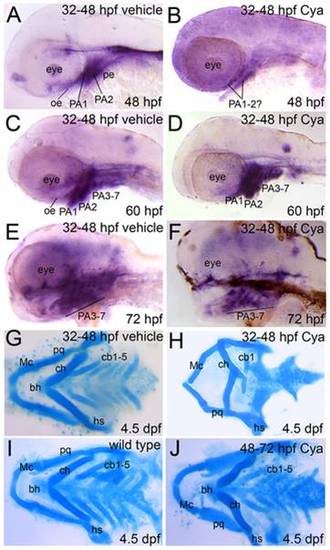
Reduced Gli activity in craniofacial tissues following late pharyngula stage Hh-pathway inhibition. Lateral (A–F) or dorsal (G–J) views, anterior to the left, of Tg(Gli-d:mCherry) fish treated with vehicle (A,C,E,G), Cya (B,D,F,H,J) or not treated (I) and stained with mCherry riboprobe (A–F) or Alcian blue solution (G–J). (A,B) Reporter gene expression was significantly reduced in the brain and facial epithelium at 48 hpf following late pharyngula stage Cya treatment. A slight mCherry signal was detected in craniofacial tissue that likely corresponds to PA1-2 mesenchyme at 48 hpf following Cya treatment. (C–F) Reporter gene expression was fully recovered in Cya treated embryos by 60–72 hpf and is expressed in oe, pe, brain and PA mesenchyme. (M–O) Alcian blue staining to reveal cartilage deficits in Gli reporter transgenic fish following Cya treatments at 32–48 hpf (H) or 48–72 hpf (J). These deficits are consistent with our previously published findings and indicate that Cya reliably inhibited cb chondrofication upon Hh-inhibition during the late pharyngula stage (H), but did not negatively impact cartilage development when administered at later stages (J). |
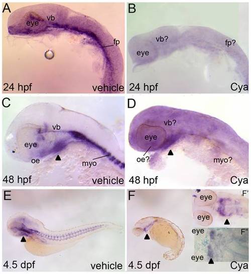
Reporter expression in Tg(Gli-d:mCherry) fish following severe reductions to Hh-signaling. Lateral (A–F) or ventral (F′,F") views, anterior to the right, of Tg(Gli-d:mCherry) fish treated with vehicle (A,C,E) or Cya (B,D,F,F′,F") and stained with mCherry riboprobe (A–F′) or dually stained with mcherry and Alcian blue (F"). Vehicle or Cya treatments began at dome stage and commenced just prior to sacrificing the embryos or larvae for staining analysis. (A,B) At 24 hpf, Cya treatments suppressed all head and trunk reporter expression in transgenic embryos. (C,D) At 48 hpf, Cya treatments reduced all reporter expression in transgenic fish, with the exception of a small expressing domain in the ventral head (arrowhead). (E,F) Reporter expression was virtually absent in Cya treated 4.5 dpf transgenic larvae, with the exception of the same mCherry expressing domain in the ventral head that was seen in 48 hpf aged embryos. (F′, F") Closer examination of the ventral head suggested that the mCherry expressing cells are within second arch mesenchyme and that no cartilage forms in this arch or any other PA in Cya treated larvae. Abbreviations: fp, floor plate; myo, myotome; vb, ventral brain. |
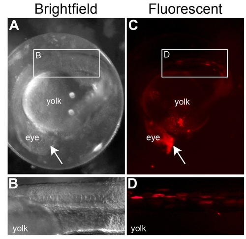
Expression of mCherry protein in Tg(Gli-d:mCherry) founder fish. (A–D) Lateral views, anterior to the left, of 24 hpf founder embryos visualized using brightfield (A,B) or fluorescent (C,D) microscopy. mCherry expressing cells could be clearly visualized in the forebrain (arrow in A,B) and myotome (boxed in A,B; see C,D for higher magnification) in some of the embryos injected with the Gli reporter transgene. |
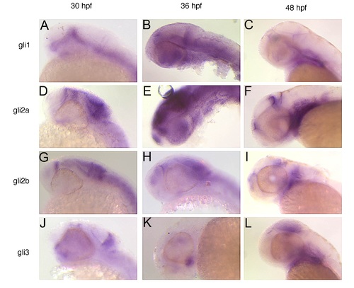
Expression of gli genes in craniofacial region during second day of development. Lateral views, anterior to the left, of 30 hpf (A,D,G,J), 36 hpf (B,E,H,K) or 48 hpf (C,F,I,L) stained with riboprobe for gli1 (A–C), gli2a (D–F), gli2b (G–I) or gli3 (J–L). |
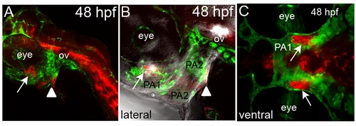
mCherry protein expression in craniofacial region of double transgenics. Lateral (A,B) or ventral (C) views of confocal stack projections of 48 hpf Tg(Gli-d:mcherry) and Fli1:gfp double transgenic fish. (A) Reporter expression was visible in craniofacial tissues. (B,C) Closer examination within the jaw showed reporter expression in the oe (arrow) and pe (arrowhead) underlying CNC within the second arch. Abbreviations: PA, pharyngeal arch; ov, otic vesicle. |
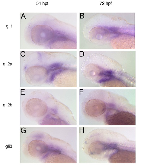
Expression of gli genes in craniofacial region during third day of development. Lateral views, anterior to the left, of 54 hpf (A,C,E,G) or 72 hpf (B,D,F,H) stained with riboprobe for gli1 (A,B), gli2a (C,D), gli2b (E,F) or gli3 (G,H). |