
Fig. 1
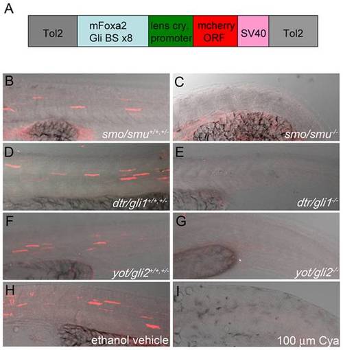
Gli-dependent reporter is capable of detecting modulations to the Hh-signaling pathway when expressed transiently in fish. (A) Schematic of the Gli-d mcherry reporter construct designed in this study. The construct consists of a 0.7 kb fragment encoding eight repeating Gli-CBS from the mouse FoxA2 floor plate enhancer and a minimal lens crystallin promoter (light blue and green respectively) inserted upstream of the mCherry ORF (red). The SV40 polyA signal (pink), which contains a transcriptional termination element, is directly downstream of the mCherry ORF and the entire element is flanked by Tol2 transposable elements (gray). (B–I) Lateral views, anterior to the left, of confocal stack projections of fixed trunk tissue. (B–G) Homozygous genetic mutants (-/-) for smu/smo (B,C), dtr/gli1 (D,E), yot/gli2 (F,G) were sorted from heterozygotes (+/-) and homozygous wild type (+/+) siblings based on evident characteristic Hh-signaling mutant phenotypes. Genetic mutants (-/-) for Hh-signaling pathway components failed to express mCherry protein (C,E,G), while strong mCherry expression was evident in trunk tissue of (+/+,+/-) siblings (B,D,F). (H,I) Hh-pathway attenuation with 100 µm Cya fully reduced mCherry protein expression in construct-expressing embryos (I) compared to vehicle-treated controls (H). |
| Fish: | |
|---|---|
| Observed In: | |
| Stage: | Prim-15 |