
- Title
-
Characterization of the AP-1 mu1A and mu1B adaptins in zebrafish (Danio rerio)
- Authors
- Zizioli, D., Forlanelli, E., Guarienti, M., Nicoli, S., Fanzani, A., Bresciani, R., Borsani, G., Preti, A., Cotelli, F., and Schu, P.
- Source
- Full text @ Dev. Dyn.
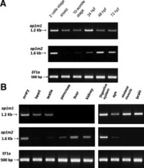
ap1m1 and ap1m2 expression during development and in adult organs. Expression analysis by RT-PCR. α-Elongation Factor served as positive control. A: ap1m1 and ap1m2 expression during zebrafish development from 2 cells to 72 hpf. B: ap1m1 and ap1m2 expression pattern in adult organs. |
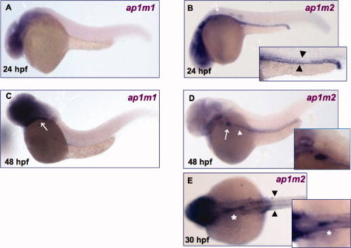
Spatio-temporal expression of ap1m1 and ap1m2. Whole mount in situ hybridization assays were performed on embryos at 24 and 48 hpf. A, C: ap1m1 expression in brain and eye at 24 hpf (A) and 48 hpf (C). Signal is also present at 48 hpf in the gut region (white arrow). B–E: ap1m2 expression. B: ap1m2 is detected at the level of pronephric ducts (black arrowhead) and gut region (white arrow) at 24 hpf. D: White arrowhead indicates digestive tract and white arrow indicates liver at 48 hpf. E: Dorsal view. Black arrowheads indicate the pronephric ducts and white asterisk indicates the intestinal bulb at 30 hpf. All embryos (except E) are mounted lateral view anterior to the left. |
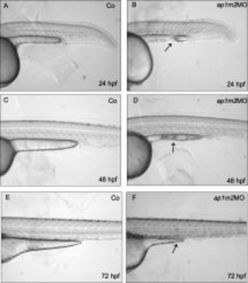
ap1m2 knock-down embryos. Morphants showed severe defects in gut tube formation and margins are not well defined. Morphants are smaller than controls. Black arrows point to particularly altered morphology. Images are at 40× magnification. Embryos are shown in lateral views, anterior to left. |
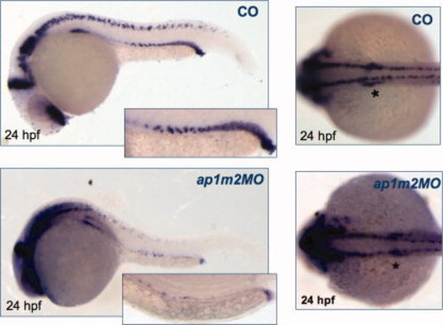
pax2a staining in control and ap1m2MO embryos. Expression of pax2a in morphants is severley reduced compared to controls at 24 hpf especially in the region of pronephric ducts (*). Embryos are shown anterior to the left and dorsal up. EXPRESSION / LABELING:
|
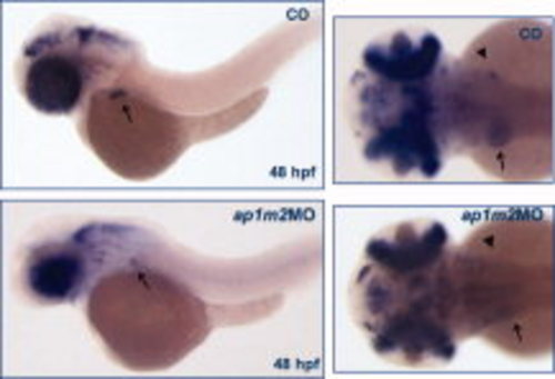
prox1 staining in control and ap1m2MO embryos. The expression of prox1 in morphants is reduced compared to controls at 48 hpf in liver (black arrows) and pancreas (black arrowheads). Embryos are shown anterior to the left and dorsal up at 25× fold magnification. EXPRESSION / LABELING:
|
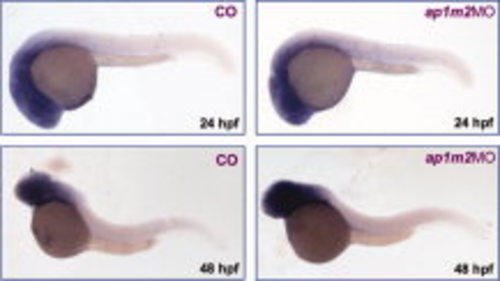
Expression level of ap1m1 transcript in ap1m2 morphants. ap1m1 expression at 24 and 48 hpf and in ap1m2 morphants. Embryos are shown anterior to the left. |
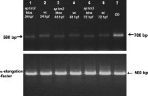
ap1m2 expression in wild type and morphants embryos. Splice-inhibiting morpholino was designed to exclude exon 3. cDNA was prepared from 24-, 48-, and 72-hpf injected-embryos and controls (see Experimental Procedures section). The expected bands were 700 bp for wild type embryos and 580 bp for injected embryos. α-Elongation-factor 1a (EF1a) serves as control. |
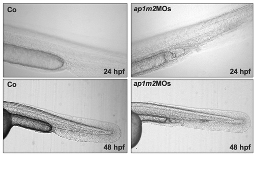
Morphants phenotype using the splice-inhibiting morfolino. Also, these morphants show severe developmental abnormalities of gut region at 24 and 48 hpf. All images are lateral view anterior to the left. |

Unillustrated author statements |