- Title
-
Identification of a heritable model of testicular germ cell tumor in the zebrafish
- Authors
- Neumann, J.C., Dovey, J.S., Chandler, G.L., Carbajal, L., and Amatruda, J.F.
- Source
- Full text @ Zebrafish
|
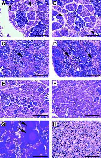
Histology of zebrafish GCTs. Hematoxylin and eosin–stained sections from formalin-fixed, paraffin-embedded zebrafish testes. (A, B) Normal testis. The testis consists of cysts or lobules of spermatogenic cells surrounded by a basement membrane and somatic cells. Small clusters of spermatogonia are seen adjacent to the basement membrane (arrowheads). Successive stages of differentiation are also evident, including primary and secondary spermatocytes, spermatids, and mature spermatozoa. (C–G) Testicular GCTs. The images shown are from the GCT mutant line; similar images can be found in carcinogen-treated fish and in males of advanced age. (C–E) Loss of orderly architecture and impaired differentiation. Ectopic clusters of spermatogonia are seen in the center of the lobule, distant from the basement membrane (arrows). In (E), a severe reduction in spermatogenesis is evident. (F) Complete loss of spermatocytic differentiation. The architecture of the lobule is overrun by a proliferation of primitive, spermatogonial-like cells. (G) Early stage I and stage II oocytes (arrows) within a testicular GCT. (H) A GCT arising on the ovary of a female after treatment with DMBA. Large, primitive-appearing germ cells with clear cytoplasm are evident (scale bar = 50μm in all images). PHENOTYPE:
|
|
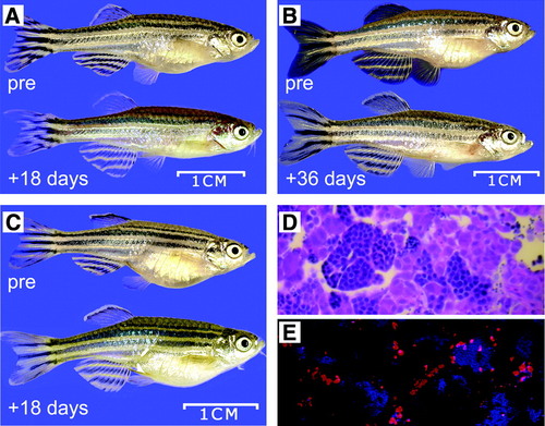
Zebrafish testicular GCTs are susceptible to radiation therapy. (A–C) Three different males that developed GCTs were irradiated (24 Gy whole-body irradiation in six daily fractions of 4 Gy). The animals were allowed to recover, and follow-up photographs were taken at the indicated time. Radiation treatment led to regression of the abdominal tumor in two cases (A, B) but had no effect on the third (C). (D) Hematoxylin and eosin image of a testicular GCT from a different fish 7 days after radiation treatment. (E) TUNEL staining of a part of the same GCT shown in (C). TUNEL-positive cells undergoing apoptosis are shown in red. Counterstaining with Hoechst 33342 (blue) indicates the condensed chromatin of spermatocytes. Apoptosis is occurring primarily in the spermatogonial-like cells. |
|
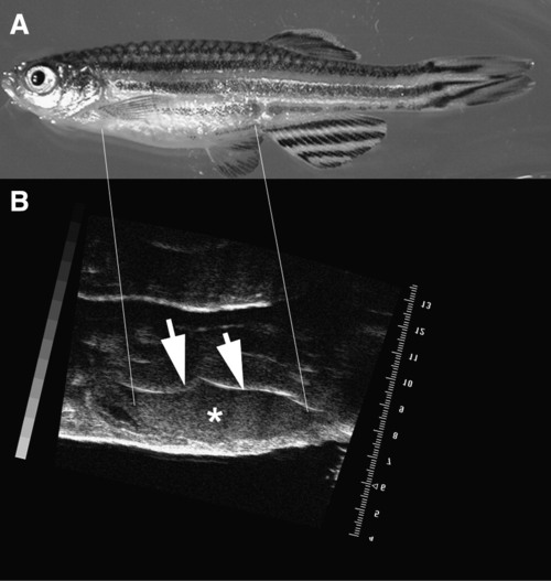
Zebrafish testicular GCTs can be detected with ultrasound. High-frequency ultrasound was used to detect a subclinical tumor in a male carrier of the TCGT susceptibility trait (A). The tumor appears as a homogenous mass (asterisk) between the ventral abdominal wall and the brightly echogenic edge of the swim bladder (arrows) (B). |


