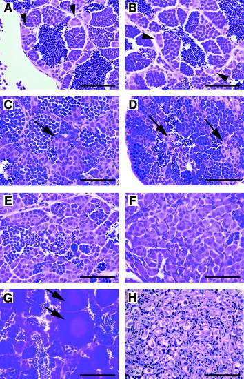
Histology of zebrafish GCTs. Hematoxylin and eosin–stained sections from formalin-fixed, paraffin-embedded zebrafish testes. (A, B) Normal testis. The testis consists of cysts or lobules of spermatogenic cells surrounded by a basement membrane and somatic cells. Small clusters of spermatogonia are seen adjacent to the basement membrane (arrowheads). Successive stages of differentiation are also evident, including primary and secondary spermatocytes, spermatids, and mature spermatozoa. (C–G) Testicular GCTs. The images shown are from the GCT mutant line; similar images can be found in carcinogen-treated fish and in males of advanced age. (C–E) Loss of orderly architecture and impaired differentiation. Ectopic clusters of spermatogonia are seen in the center of the lobule, distant from the basement membrane (arrows). In (E), a severe reduction in spermatogenesis is evident. (F) Complete loss of spermatocytic differentiation. The architecture of the lobule is overrun by a proliferation of primitive, spermatogonial-like cells. (G) Early stage I and stage II oocytes (arrows) within a testicular GCT. (H) A GCT arising on the ovary of a female after treatment with DMBA. Large, primitive-appearing germ cells with clear cytoplasm are evident (scale bar = 50μm in all images).
|
