FIGURE SUMMARY
Title

Genetic interaction of PGE2 and Wnt signaling regulates developmental specification of stem cells and regeneration

Authors
Goessling, W., North, T.E., Loewer, S., Lord, A.M., Lee, S., Stoick-Cooper, C.L., Weidinger, G., Puder, M., Daley, G.Q., Moon, R.T., and Zon, L.I.
Source
Full text @ Cell

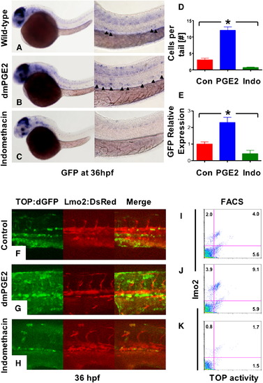
Prostaglandin Levels Directly Affect wnt Activity in Zebrafish Embryos

(A–C) In situ hybridization for GFP in TOP:dGFP wnt reporter embryos at 36 hpf shows widespread wnt activity; inset, close-up of GFP+ (black arrowheads) cells in the AGM. 10 μM dmPGE2 enhanced GFP expression throughout the embryo, while 10 μM indo decreased global wnt activity, and in the AGM.

(D) Quantification (mean ± SD) of total GFP+ cells in the major trunk vessels and (E) qPCR analysis for GFP in whole embryo lysates following exposure to dmPGE2 or indo versus vehicle con reveals significant alterations in wnt activity (*significant (sig) across treatment groups, ANOVA, p < 0.05, n = 10/treatment).

(F–H) Representative confocal microscopy images of the AGM region of TOP:dGFP; lmo2:DsRed embryos following exposure to con, dmPGE2, or indo are shown; differences can be seen in the wnt-active (green, left column), HSC/endothelial (red, middle), and colocalized (merged, right) populations.

(I–K) Representative FL1 (green)/ FL2 (red) FACS plots of individual TOP:dGFP; lmo2:DsRed embryos after exposure to con, dmPGE2 or indo confirm the confocal analyses (summarized in Figure S2).

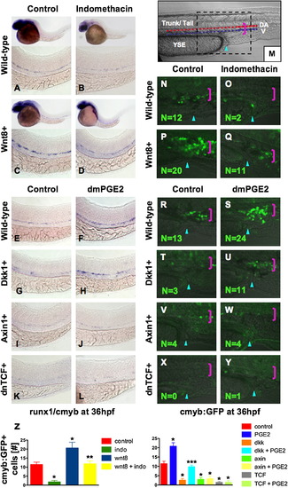
PGE2 Regulates Wnt-Mediated Effects on HSC Formation at the Level of β-catenin

(A–D) wnt8 induction in hs:wnt8-GFP transgenic embryos increased runx1/cmyb+ HSCs compared to WT; indo decreased HSCs in WT and wnt8 embryos.

(E–L) Induction of dkk diminished runx1/cmyb expression compared to WT. dmPGE2 enhanced HSCs in WT embryos, and rescued runx1/cmyb expression to approximately WT levels in dkk embryos. Axin (hs:axin-GFP) and dnTCF (hs:dnTCF-GFP) reduced runx1/cmyb expression, and dmPGE2 could not rescue those effects.

(M) Schematic of confocal microscopy. Imaging was performed in the trunk/tail region of the embryo, centered around the tip of the yolk sac extension (YSE, blue arrowhead), encompassing the dorsal aorta (red dots) and vein (blue dots), as indicated by the pink bracket.

(N-Y) In vivo confocal microscopy of wnt pathway inducible embryos crossed into the cmyb:GFP HSC reporter line confirmed the in situ hybridization analysis and demonstrated quantifiable effects on cell number.

(Z) Cell counts (5 embryos/treatment, data represented as mean ± SD) were conducted in the major vessels (pink bracket) in a 40x field centered at the YSE; *sig versus con; **sig versus wnt8; ***sig versus dkk; ANOVA, p < 0.001.

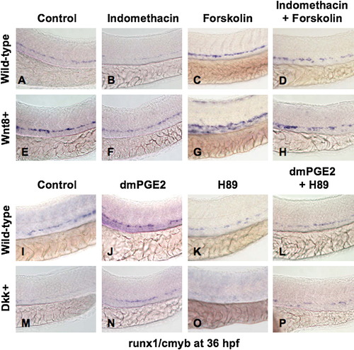
PGE2 Regulates the Effects of wnt Activity on HSCs via cAMP/PKA Signaling

(A, B, E, F, I, J, M, and N) The /PGE2/wnt interaction affected runx1/cmyb+ HSC formation, as seen in Figure 2. (C and G) cAMP enhancement by forskolin increased runx1/cmyb expression in WT embryos and further expanded HSCs in wnt8 embryos. (D and H) Forskolin counteracted the inhibitory effect of indo on HSC formation in WT and wnt8 embryos. (K and O) PKA inhibition by H89 decreased runx1/cmyb expression in WT embryos and eradicated HSC formation in dkk embryos. (L and P) The enhancing effect of dmPGE2 on HSCs was reduced back to baseline levels by PKA inhibition; similarly, the dmPGE2-induced rescue of HSCs in dkk embryos was blocked by H89.

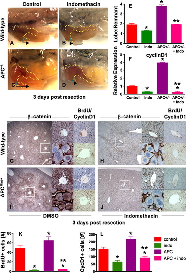
The PGE2/wnt Interaction Is a Master Regulator of Liver Regeneration

(A–D) Representative photomicrographs of en bloc dissections following liver resections at day 3 are shown; the liver is highlighted by a yellow dotted line, the resection site by a blue line, and the black arrow indicates the amount of liver regrowth. Wnt activation in APC mutant zebrafish enhanced liver regeneration compared to WT. Indo stymied liver regeneration.

(E) Quantification of zebrafish liver regeneration showed significant differences across treatment groups; *sig versus con, **sig versus APC+/-, ANOVA, p < 0.001, n ≥ 6.

(F) qPCR for cyclin D1 gene expression; effects are coordinately regulated by the PGE2/wnt interaction; *sig versus con, **sig versus APC, ANOVA, p < 0.001, n = 7.

(G–J) wnt and PGE2 modulation has significant effects on murine liver regeneration following 2/3 partial hepatectomy. APCMin mice exhibit enhanced β-catenin staining (left panel), particularly in the periportal areas (top middle panel), with noticeable nuclear staining (bottom middle panel). BrdU incorporation (top right panel) and cyclin D1 staining (bottom right panel) indicated enhanced regenerative activity. Indo diminished global β-catenin staining (left and top middle panels), excluded β-catenin from the nuclei (bottom middle panels), and resulted in a corresponding decrease of both BrdU incorporation and cyclin D1.

(K and L) Quantification of BrdU incorporation and cyclinD1 staining in corresponding serial sections of regenerating livers; *sig versus con, **sig versus APC, ANOVA, p < 0.05, n = 10 sections/treatment.

Data represented as mean ± SD.

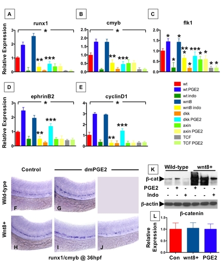
PGE2 affects the expression of HSC and vascular markers by increasing β-catenin levels
Embryos were heat shocked at 5 somites and then exposed to DMSO, 10μM dmPGE2, or 10μM indomethacin as indicated from 10 somites until 36 hpf.
(A – E) Quantitative PCR was performed on cDNA obtained from whole embryo extracts. The primers are listed in Supplementary Table 1. An asterisk (*) indicates a statistically significant difference compared to the wild-type control, ANOVA, p<0.001, n≥5 sets of 50 embryos/condition; data shown as mean ± SD.
(A and B) wnt and PGE2 pathway modulation affected the expression of HSC markers runx1 and cmyb.
(C and D) wnt and PGE2 pathway modification altered the expression of the vascular marker flk1 and arterial marker ephrin B2.
(E) The wnt target cyclin D1 was also affected by the interaction of wnt and PGE2 signaling.
(F - J) wnt8 induction and dmPGE2 treatment synergistic regulated runx1/cmyb+ HSCs; exposure of wild-type embryos to dmPGE2 enhanced HSC number (G), as did induction of wnt8 (H). Combinatorial treatment (I) lead to increased runx1/cmyb staining in 25/41 embryos. (J) In 5/41 embryos, runx1/cmyb expression was reduced compared to wildtype after combined treatment with wnt8 and dmPGE2, indicating a potential negative effect of overactivation of the wnt pathway on HSC formation.
(K) Western blot analysis of total β-catenin in zebrafish embryo homogenates (12 embryos/lane) at 36 hpf. Induction of wnt8 as well as dmPGE2 treatment increased β- catenin, while indomethacin decreased levels in both wild-type and wnt8 embryos.
(L) qPCR for β-catenin revealed a lack of transcriptional upregulation following wnt8 induction or dmPGE2 treatment (ANOVA, p=0.937, n=6 sets of 50 embryos/condition).

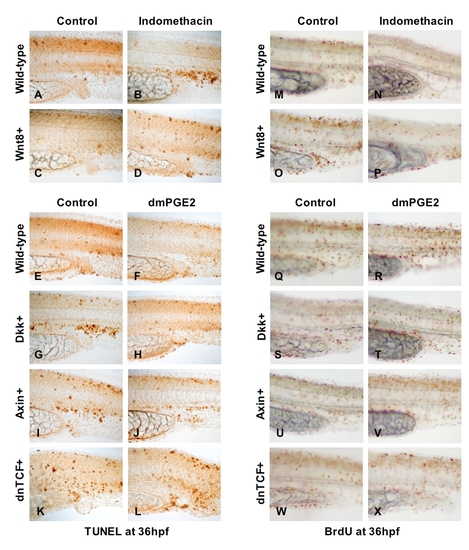
PGE2-mediated modulation of wnt signaling affects cell death and proliferation
(A – D) Indo treatment enhanced TUNEL+ cells in the AGM in both WT and wnt8 induced embryos; median representatives are shown.
(E – L) Induction of dkk, axin, or dnTCF enhanced apoptosis; dmPGE2 improved this effect only in dkk transgenics.
(M – P) Induction of wnt8 enhanced BrdU incorporation, while indo treatment diminished BrdU in both WT and wnt8 embryos.
(Q – X) Inhibition of wnt signaling by dkk, axin, or dnTCF diminished BrdU incorporation; the effect of dkk could be rescued by dmPGE2.

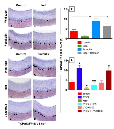
PGE2 enhances wnt activity in the HSC compartment via the PKA/cAMP pathway
(A – D, F - K) In situ hybridization for GFP in TOP:dGFP wnt reporter embryos following treatment with modifiers of PGE2/cAMP/PKA-mediated signaling at 36 hpf.
(A and C) cAMP stimulation by forskolin (0.5μM) enhanced the number of wnt active cells within the tail region of the zebrafish embryo.
(B and D) Indomethacin (10μM) decreased wnt activity in the zebrafish embryo. This effect could be rescued by the addition of forskolin.
(E) Quantitative summary of the effects of indomethacin and forskolin on TOP:dGFP+ cells in the AGM region. * statistically significant between each other, ANOVA, p< 0.001, n=10).
(F and G) Exposure to dmPGE2 enhanced GFP expression in the tail region.
(H and I) PKA inhibition by H89 (0.5μM) decreased wnt activity in wild-type embryos and diminished the wnt-enhancing effects of dmPGE2.
(J and K) Inhibition of PI3K by LY294002 (1μM) had no effect on wnt activity in wildtype and dmPGE2-treated embryos.
(L) Summary of the effects of dmPGE2, H89, and LY294002 on TOP:dGFP+ cells in the AGM region. * statistically significant between each other, ANOVA, p<0.001, n=10).

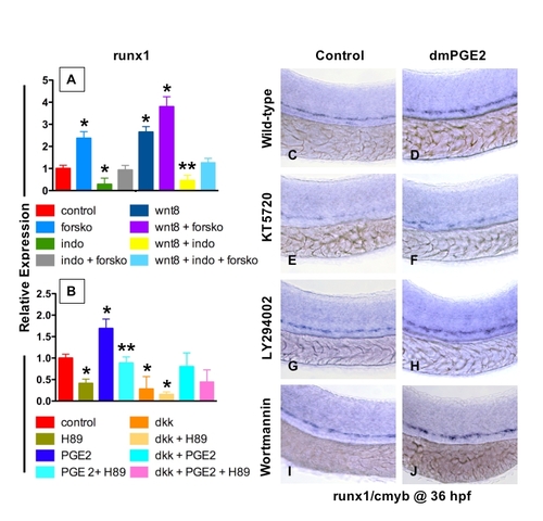
PGE2 enhances wnt activity in the HSC compartment via the PKA/cAMP pathway
(A and B) qPCR results for runx1 expression after wnt8 transgene induction and chemical treatments. * statistically significant vs. control; ** statistically significant vs. wnt8 (A) or dmPGE2 (B), ANOVA, p<0.001; n=3 sets of 50 embryos; data represented as mean ± SD.
(A) cAMP stimulation by forskolin (0.5μM) and wnt8 induction enhanced runx1 levels at 36 hpf in qPCR analysis of whole embryo lysates. Forskolin improved the negative effects of indomethacin treatment in both controls and wnt8 transgenics.
(B) PKA inhibition by H89 (0.5μM) reduced runx1 expression by qPCR in both control and dmPGE2 treated embryos.
(C and D) dmPGE2 (10μM) enhanced runx1/cmyb expression in the zebrafish tail at 36 hpf.
(E and F) PKA inhibition by KT5720 (1μM) had similar effects on HSC number in wildtype and dmPGE2-treated embryos as seen with H89 exposure.
(G and H) PI3K inhibition by LY294002 (1μM) or (I and J) Wortmannin (1μM) did not affect HSC number in wild-type or dmPGE2-treated embryos.

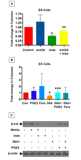
The PGE2/wnt interaction affects hematopoietic colony formation in murine ES cells
(A and B) ES cell hematopoietic colony formation assays were performed; cells were exposed to soluble modulators of the wnt pathway (10 ng/ml Wnt3A of 400 ng/ml Dkk1) and then incubated with indomethacin (20 μM), dmPGE2 (10μM), or forskolin (5 μM) as indicated. Colony forming units-Culture (CFU-E (erythroid), CFU-M (monocyte), CFUG (granulocyte), CFU-GM (granulocyte-monocyte), and CFU-GEMM (granulocyteerythroid- monocyte-megakaryocyte)) were scored on day 8-10 based on morphology. Averages ± SEM in the fold changes in total CFU-C number of each sample relative to control were calculated from at least six individual experiments.
(A) The positive effect of wnt activation on hematopoietic differentiation could be diminished by inhibition of prostaglandin synthesis with indomethacin. Wnt3A enhanced hematopoietic colony formation. Indomethacin (20μM) alone diminished colony number and inhibited the enhancing effects of Wnt3A (* statistically significant vs. control, t-test, p=0.002, ** statistically significant vs. indomethacin, p<0.001; n=6).
(B) Inhibition of wnt activity decreased hematopoietic differentiation and could be rescued by PGE2 or cAMP signaling. Inhibition of wnt activation by Dkk1 (10ng/ml) diminished colony number. dmPGE2 (10μM) and the cAMP activator forskolin (0.5μM) enhanced hematopoietic colonies. This negative effect of Dkk1 could be rescued by both dmPGE2 or forskolin, confirming the role of PKA in this process. (* statistically significant vs. control, t-test, p=0.005, *** statistically significant vs. Dkk1, p<0.046; n=4).
(C) Western blot analysis of ES cell lysates. Wnt3A and dmPGE2 treatment enhanced β- catenin levels, while indomethacin and Dkk1 had negative effects. Indomethacin diminished the enhancing effect of Wnt3A, whild dmPGE2 could enhance β-catenin levels after Dkk1 exposure.

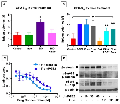
PGE2 affects hematopoietic progenitor function by enhanced cAMP production and subsequent phosphorylation events
(A – B) CFU-S12 colony formation after transplantation of 500 purified KSL cells into irradiated recipient C57Bl/6 mice. Spleens were dissected at day 12 after transplantation and colonies counted.
(A) Effect of in vivo treatment of lethally irradiated recipient mice with indomethacin (2.5 mg/kg, i.p., q.o.d.), BIO (50μg/kg), or both, on CFU-S12 colony formation; BIO treatment significantly increased colony formation, which could be inhibited by concurrent treatment with indomethacin (* statistically different from control and other treatments, t-test, p=0.015, n≥7).
(B) Effect of ex vivo treatment of purified KSL cells with Dkk1 (10ng/ml), dmPGE2 (10μM), forskolin (Fors; 1μM), cholera toxin (Chol Tox; 0.5μM) and/or a combination of drugs as indicated on CFUS12 activity. Inhibition of wnt activity by Dkk1 reduced colony formation. This effect could be rescued by both dmPGE2 and direct cAMP stimulation by forskolin or cholera toxin; * statistically significant vs. control, ANOVA, p<0.05, n=7- 10.
(C) cAMP luminescence assay in isolated murine bone marrow cells; luminescence intensity is inversely related to cAMP levels. Exposure of bone marrow with dmPGE2 and forskolin for 15 minutes had dose-dependent enhancing effects on cAMP levels (mean ± SD, n=3).
(D) Western blot analysis of homogenized murine bone marrow cells following ex vivo drug treatment. Total β-catenin levels increased in a time-dependent fashion following exposure to dmPGE2 (10μM), and decreased in response to indomethacin (10μM). The response of phospho-β-catenin(S675) and phospho-GSKβ(S9) to drug treatment was also time dependent, and preceded effects on total β-catenin.

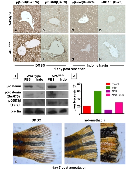
The interaction of PGE2 and wnt affects regeneration in several organs (A - H) Immunohistochemical analysis of liver samples 1 day post 2/3 partial hepatectomy in C57Bl/6 and APCMin mice. Photomicrographs were taken at 20x magnification.
(A and E) Heterozygous APC loss enhanced site-specific phosphorylation of β-catenin (S675) post liver resection.
(B and F) Heterozygous APC loss enhanced site-specific phosphorylation of phospho- GSK3β (S9). (C, D, G, H) Inhibition of prostaglandin synthesis with indomethacin (2.5 mg/kg twice daily intraperitoneal injections) resulted in undetectable levels of phospho-β-catenin (S675) and phospho-GSK3β (S9).
(I) Inhibition of PGE2 production by indomethacin diminished total β-catenin levels and site-specific phosphorylation of β-catenin (S675) and GSK3β (S9) in mice following partial hepatectomy by western blot analysis.
(J) Inhibition of PGE2 synthesis increased hepatocellular necrosis in regenerating mouse livers. Histological sections of mouse livers were examined for extensive areas of necrosis. Sections with more than 4 areas of necrosis were counted as positive. Indomethacin increased areas of necrosis in wild-type and APCMin mice.
(K and L) Effect of PGE2 inhibition on zebrafish fin regeneration.
(K) Following amputation, the zebrafish caudal fin regenerated completely over 7 days through a wnt-mediated process of progenitor proliferation (blastema).
(L) Treatment with indomethacin (20μM) completely blocked the regenerative process compared with DMSO-treated controls (n=3/3).

Acknowledgments
This image is the copyrighted work of the attributed author or publisher, and ZFIN has permission only to display this image to its users. Additional permissions should be obtained from the applicable author or publisher of the image.

Reprinted from Cell, 136(6), Goessling, W., North, T.E., Loewer, S., Lord, A.M., Lee, S., Stoick-Cooper, C.L., Weidinger, G., Puder, M., Daley, G.Q., Moon, R.T., and Zon, L.I., Genetic interaction of PGE2 and Wnt signaling regulates developmental specification of stem cells and regeneration, 1136-1147, Copyright (2009) with permission from Elsevier. Full text @ Cell