
- Title
-
Glycogen synthase kinase 3alpha and 3beta have distinct functions during cardiogenesis of zebrafish embryo
- Authors
- Lee, H.C., Tsai, J.N., Liao, P.Y., Tsai, W.Y., Lin, K.Y., Chuang, C.C., Sun, C.K., Chang, W.C., and Tsai, H.J.
- Source
- Full text @ BMC Dev. Biol.
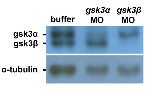
Injection of translation inhibitors gsk3α- and gsk3β -MO into embryos can specifically reduce the protein levels of GSK3α and GSK3β, respectively. The total protein lysate extracted from seven zebrafish embryos at 24 hpf was loaded on each lane and analyzed by western blot. The antibody used is indicated in the left of each blot. Anti-GSK3 antibody enables to recognize both GSK3α and GSK3β proteins; anti-α-tubulin antibody was used as a loading control. The protein levels of GSK3α and GSK3β were reduced greatly in the protein lysates extracted from the gsk3α – and gsk3β -MO-injected embryos, respectively. |
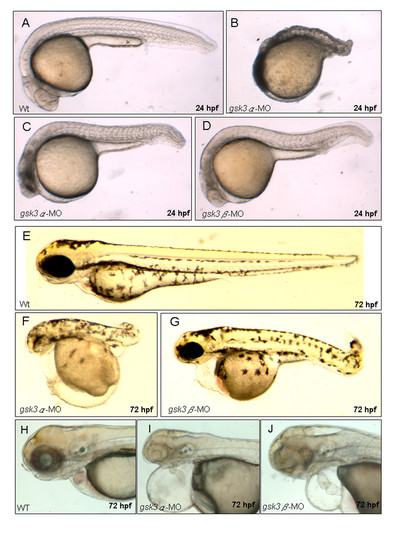
The morphological defects in gsk3α and gsk3β morphants. Wild-type embryos (A, E, H), gsk3α (B, C, F, I), and gsk3β (D, G, J) morphants. The 24 hpf gsk3α morphants have mild (C) to severe (B) defects in axis formation. At 72 hpf, both gsk3α and gsk3β morphants displayed pericardial edema (F, G, I, J) and an unlooped, stretched heart (I, J). PHENOTYPE:
|
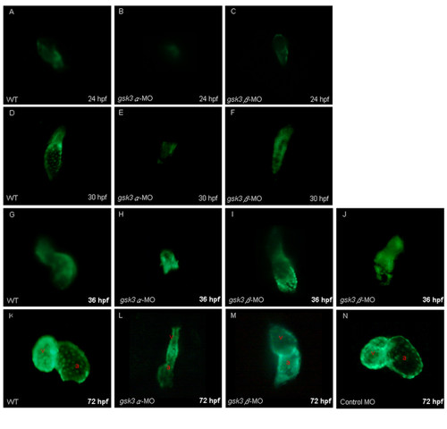
The cardiac defects induced by the knockdown of zebrafish GSK3α and GSK3β. Anti-sense morpholino oligonucleotide (MO), which was designed to specifically inhibit the translation of either gsk3α-(gsk3α-MO) or gsk3β-mRNA (gsk3β-MO), was injected into one-celled stage embryos and the heart morphology was observed at the stage as indicated. The elongation of heart tube was normally developed at 24 hpf in the wild-type (A) and in the gsk3β morphants (C); whereas the heart of gsk3α morphant did not elongate to from a heart-tube (B). The wild-type (D) and gsk3β morphant's heart (F) developed normally at 30 hpf, but the heart of gsk3α morphant was still retardant development at 30 hpf (E), and even ceased at heart-cone stage at 36 hpf (F). Compared to the wild-type (G), however, the heart positioning was abnormally in the gsk3β morphant at 36 hpf (I, J). Eventually, both gsk3α and gsk3β morphants displayed an unlooped and stretched heart (L, M). The heart morphology of embryos injected with the control MO was also observed at 72 hpf (N). a: atrium; v: ventricle. |
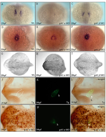
The heart defects in gsk3α morphants weredue to a reduced cardiomyocyte population size. Dorsal (A-I) and lateral (J-O) views of embryos stained by in situ hybridization (A-F) and TUNEL assay (G-L). Whole mount in situ hybridization staining with cmlc2 at 18 and 23 hpf received that gsk3α-MO causes a repressive influence on cardiomyocyte formation (B, E). The heart defect in gsk3α morphants was due to the reduction of cardiomyocyte population size. However, gsk3β morphants display normal cardiomyocyte formation (C, F) compared to wild-type embryos (A, D). TUNEL labeling was evident throughout the head of gsk3a-MO-injected embryos (H), especially in the head, but was limited in the head of controls (G) and gsk3β morphants (I). Compared to embryos derived from the transgenic line, Tg(cmlc2:EGFP), which has heart-specific GFP (K), we observed that the heart of gsk3α morphant did not elongate to form a heart-tube and the GFP signal was very faint at 24 hpf (N). Panels L and O are the merged images from J and K, and M and N, respectively. The apoptotic signals were co-localized with the heart-specific GFP signal, indicating that the reduced cardiomyocyte numbers was due to apoptosis in heart (O). h: heart. EXPRESSION / LABELING:
PHENOTYPE:
|
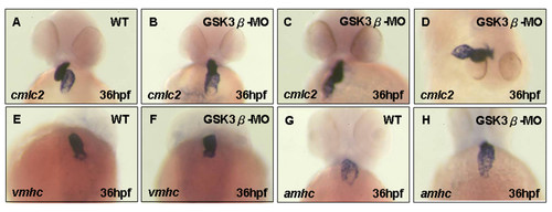
Cardiac positioning is gsk3β-dependent, but the chamber-specific patterning is not. A-C, E-H) ventral view, (D) dorsal view of wild-type (A, E, G), and gskβ morphants (B-D, F, H) at 36 hpf. in situ hybridization with cmlc2 staining revealed that randomized looping was observed in gsk3β morphants (B-D). The expression of vmhc (F) and amhc (H) appeared normal in gsk3β morphants. |
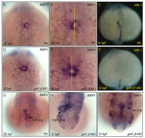
Heart asymmetry was affected in gsk3β morphants. Normally, bmp4 transcripts accumulate predominantly on the left side of the heart tube at 20 hpf (A, B), and the left-predominant bmp4 asymmetry persists through the stages of jogging (G). However, in gsk3β morphants, the expression of bmp4 becomes symmetrical at 20 hpf (B, D). In gsk3β morphants, in which the heart fails to jog, bmp4 is more evenly distributed in the heart region (H, I). The left-sided lefty-1 domain was greatly reduced in gsk3β morphant hearts at 16 hpf (F). All are dorsal views. B, E are higher magnifications of A, D, respectively. Lines mark the midline. L, embryo left. EXPRESSION / LABELING:
PHENOTYPE:
|
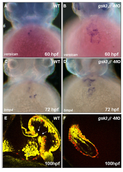
GSK3β modulates zebrafish cardiac valve formation. Whole-mount in situ hybridization with bmp4 and versican staining reveals that cardiac valve development was affected in gsk3β morphants. At 60–72 hpf,versican (A, B) and bmp4 (C, D) expression was greatly up-regulated in gsk3β morphants. Tg(cmlc2: Hc-RFP) embryos were injected with gsk3β-MO and observed by in vivo two-photon fluorescence imaging of a live transgenic zebrafish heart at 100 hpf. The endocardial cells and blood are labeled yellow; the Hc-GFP-positive myocardial cells are labeled red. Valves are clearly observed in wild-type embryos (E; white arrows), but not in gsk3β morphants (F). b, blood cells; V, ventricle; A, atrium. EXPRESSION / LABELING:
PHENOTYPE:
|
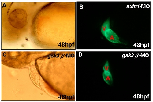
Similar cardiac defects in the axin1 and the gsk3β morphants. Axin1-MO or gsk3β-MO were microinjected to and observed under dissecting microscope by bright field (A, C) or fluorescence (B, D). Incomplete looping of the heart tube was also observed in axin1 mutant heart. PHENOTYPE:
|
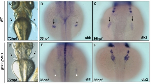
Arrested pectoral fin bud induction in gsk3β morphants. At 72 hpf, wild-type pectoral fins elongate (A), but gsk3β morphants have still not developed fin buds (arrows; D). Whole mount in situ hybridization with shh and dlx2 staining reveal that the developed of fin bud were affected in gsk3β morphants. At 36 hpf, wild-type embryos continue shh (B) and dlx2 (C) expression in the developing bud mesenchyme, but in gsk3β morphants, the shh and dlx2 expression is greatly decreased. EXPRESSION / LABELING:
PHENOTYPE:
|