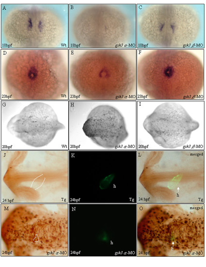
The heart defects in gsk3α morphants weredue to a reduced cardiomyocyte population size. Dorsal (A-I) and lateral (J-O) views of embryos stained by in situ hybridization (A-F) and TUNEL assay (G-L). Whole mount in situ hybridization staining with cmlc2 at 18 and 23 hpf received that gsk3α-MO causes a repressive influence on cardiomyocyte formation (B, E). The heart defect in gsk3α morphants was due to the reduction of cardiomyocyte population size. However, gsk3β morphants display normal cardiomyocyte formation (C, F) compared to wild-type embryos (A, D). TUNEL labeling was evident throughout the head of gsk3a-MO-injected embryos (H), especially in the head, but was limited in the head of controls (G) and gsk3β morphants (I). Compared to embryos derived from the transgenic line, Tg(cmlc2:EGFP), which has heart-specific GFP (K), we observed that the heart of gsk3α morphant did not elongate to form a heart-tube and the GFP signal was very faint at 24 hpf (N). Panels L and O are the merged images from J and K, and M and N, respectively. The apoptotic signals were co-localized with the heart-specific GFP signal, indicating that the reduced cardiomyocyte numbers was due to apoptosis in heart (O). h: heart.
|
