- Title
-
Bruno-like protein is localized to zebrafish germ plasm during the early cleavage stages
- Authors
- Hashimoto, Y., Suzuki, H., Kageyama, Y., Yasuda, K., and Inoue, K.
- Source
- Full text @ Gene Expr. Patterns
|
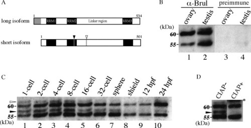
Western Blot Analysis of zebrafish Bruno-like (Brul) protein. (A) Schematic representation of the two isoforms of Brul proteins. Both forms possess three RNA recognition motifs (RRMs). The long isoform (GenBank Acc. No.BC049453) contains an additional 38 amino acid residues in its N-terminal region (gray box), whereas the short isoform has one extra amino acid residue in the RRM2 region (filled arrowhead), and four in the linker region (open arrowhead; GenBank Acc. No. AB032726), probably due to alternative splicing. (B) Two isoforms of Brul (approximately 55 and 60 kDa) are expressed in both male and female gonads. 50 μg of protein was loaded in each lane. The 60-kDa isoform is abundant in the ovary (lane 1), whereas no difference in the amounts of the two isoforms was observed in the testis (lane 2). No significant band was detected in the Western Blot with preimmune serum (lanes 3, 4). (C) Major two isoforms of Brul protein were observed throughout embryogenesis. Two phosphorylated proteins (approximately 57 kDa, filled arrowhead; and 62 kDa, open arrowhead) were also observed during early embryogenesis. Signal intensity at the shield and 12-hpf (6-somite) stages was relatively weak, consistent with faint signals in whole-mount immunostaining (see Fig. 2). Note that from the 32-cell to 12-hpf stages, the 55-kDa band is stronger than that at 60 kDa, whereas the 60-kDa band is stronger at the 24 hpf stage (lane 10). (D) Slower migrating bands disappear after CIAP treatment. Protein from blastomeres of 8-cell stage embryos was incubated at 37 °C in the presence or absence of CIAP. The 57-kDa protein, in particular, was almost completely eliminated. The asterisk represents CIAP protein. |
|
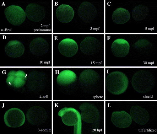
Stereomicroscopic observation of Brul expression after whole-mount immunostaining. In each panel the embryo on the left was immunoreacted with anti-Brul antiserum, and that on the right was reacted with preimmune serum as negative control. Brul is distributed maternally (A–C), accumulates at the animal pole during the 1-cell stage (D–F), and exists over the whole embryos throughout embryonic development (G–K). During early cleavage stages, specific accumulation was observed at the ends of the cleavage furrows (arrowheads in G), whereas Brul was distributed evenly in the blastomeres (G). Fluorescent intensity was reduced in the shield and 3-somite stages. Brul was also detected in unfertilized embryos (L). A–F and L are lateral views with the animal pole up; G and H are lateral oblique views; I is an animal pole view with dorsal side left; J and K are lateral views. |
|
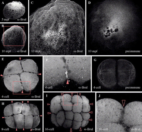
Confocal microscopic analysis of Brul protein during the early cleavage stages. Ubiquitous distribution of Brul is also detected in the blastomeres. A large number of minute particles are distributed throughout the cortex (B and C). No signal was observed inside the yolk. These particles were detected after 10 min post-fertilization (mpf) but not in either 5 mpf embryos (A) or control embryos with preimmune serum (D). In the 4-cell stage embryos, strong accumulation of Brul was detected at the ends of the cleavage furrows (arrowheads is E). In addition to this accumulation, numerous tiny dots were distributed along the marginal regions of the blastomeres (E, F). No significant fluorescence was observed in embryos stained with preimmune serum (G). A similar pattern of localization was observed in the 8-cell stage (H, arrowheads). In the 16-cell stage embryos, however, neither the accumulation at the ends of the cleavage furrows nor the tiny dots at the marginal regions were found (I; open arrowheads represent the ends of the cleavage furrows without Brul protein). C, F and J represent higher magnifications of rectangles in B, E and I, respectively. E–J show animal pole views of the embryos. |
Reprinted from Gene expression patterns : GEP, 6(2), Hashimoto, Y., Suzuki, H., Kageyama, Y., Yasuda, K., and Inoue, K., Bruno-like protein is localized to zebrafish germ plasm during the early cleavage stages, 201-205, Copyright (2006) with permission from Elsevier. Full text @ Gene Expr. Patterns


