
|
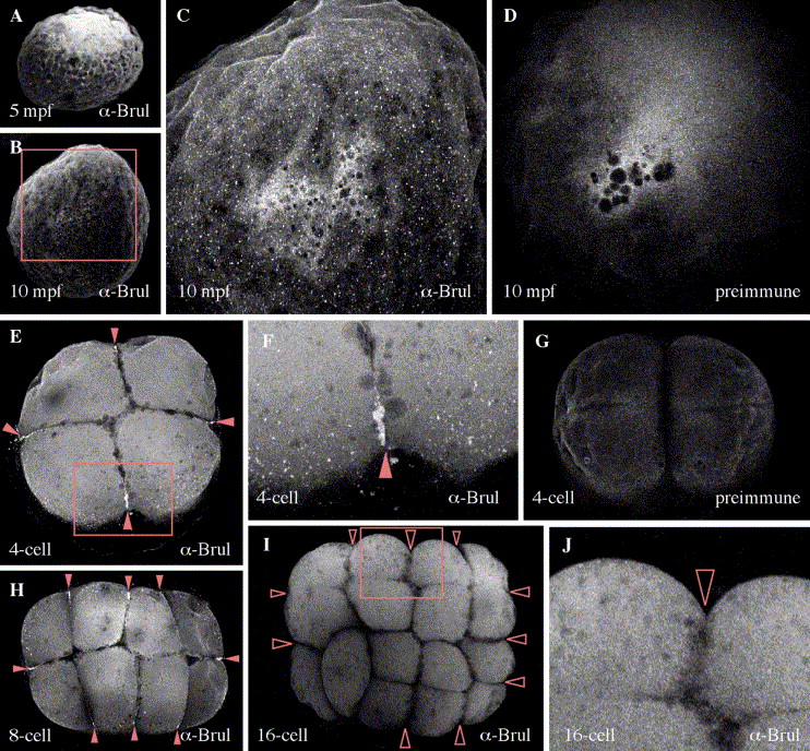
Fig. 3 Confocal microscopic analysis of Brul protein during the early cleavage stages. Ubiquitous distribution of Brul is also detected in the blastomeres. A large number of minute particles are distributed throughout the cortex (B and C). No signal was observed inside the yolk. These particles were detected after 10 min post-fertilization (mpf) but not in either 5 mpf embryos (A) or control embryos with preimmune serum (D). In the 4-cell stage embryos, strong accumulation of Brul was detected at the ends of the cleavage furrows (arrowheads is E). In addition to this accumulation, numerous tiny dots were distributed along the marginal regions of the blastomeres (E, F). No significant fluorescence was observed in embryos stained with preimmune serum (G). A similar pattern of localization was observed in the 8-cell stage (H, arrowheads). In the 16-cell stage embryos, however, neither the accumulation at the ends of the cleavage furrows nor the tiny dots at the marginal regions were found (I; open arrowheads represent the ends of the cleavage furrows without Brul protein). C, F and J represent higher magnifications of rectangles in B, E and I, respectively. E–J show animal pole views of the embryos.
Reprinted from Gene expression patterns : GEP, 6(2), Hashimoto, Y., Suzuki, H., Kageyama, Y., Yasuda, K., and Inoue, K., Bruno-like protein is localized to zebrafish germ plasm during the early cleavage stages, 201-205, Copyright (2006) with permission from Elsevier. Full text @ Gene Expr. Patterns