
- Title
-
Zebrafish GADD45{beta} genes are involved in somite segmentation
- Authors
- Kawahara, A., Che, Y.S., Hanaoka, R., Takeda, H., and Dawid, I.B.
- Source
- Full text @ Proc. Natl. Acad. Sci. USA
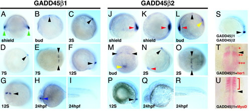
Expression patterns of zebrafish gadd45β genes. Whole-mount in situ hybridization with probes for gadd45β1 (A?I) and gadd45β2 (J?R) is shown. Stages are indicated at the lower left of each micrograph. S, somite; hpf, hours postfertilization. Lateral view, dorsal is right (A and J) and anterior is left (B?D, F, H, I, L, N, and P?S). Dorsal view, anterior is left (E and O) and anterior is up (U). Anterior view, dorsal is right (K). Posterior view, dorsal is up (G, M, and T). Expression of gadd45β1 in the YSL at the shield stage is shown in A (green arrowhead). gadd45β2 expression in the shield and involuting mesoderm is shown in J?L (red arrowhead). At bud stage, one bilateral stripe arises for both gadd45β1 and gadd45β2 at the anterior edge of presomitic mesoderm (C, L, and M; black arrowhead); weak expression of gadd45β2 is observed in the tail-bud mesoderm (L and M; yellow arrowhead). Expression in anterior presomitic mesoderm is maintained throughout somitogenesis (C?G and L?P; black arrowhead). Weak expression of gadd45β2, but not gadd45β1, is maintained in the somites. At 24 hpf, gadd45β1 expression is newly detected in the lens of the eye (H, blue arrowhead). (S) Whole-mount in situ hybridization with both gadd45β1 and gadd45β2 probes shows coexpression in the anterior PSM (black arrowhead). (T) Double in situ hybridization shows that gadd45β1 (purple and black arrowhead) is located just anterior of the second her1 expression domain (red and red asterisks) in the anterior PSM. (U) Double in situ hybridization shows that gadd45β1 is positioned posterior to the MyoD expression domain (red and red bracket), leaving a gap between the domains. |
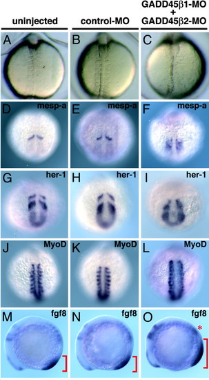
Effect of gadd45β MOs on somite segmentation. MOs (control-MO, 15 ng; GADD45β1-MO + GADD45β2-MO, 7.5 ng each) were injected into one- or two-cell-stage embryos. Dorsal view, anterior is up (A?C and J?L). Posterior view, dorsal is up (D?I). Lateral view, anterior is left (M?O). (A?C) Live embryos at 8- or 10-somite stages. Injection of GADD45β1-MO plus GADD45β2-MO, but not control-MO, caused severe defects in somite segmentation (A?C). (D?O) Whole-mount in situ hybridization with probes are shown at the upper right of each micrograph. Expression of mesp-a and her1 in the anterior PSM and segmental expression of MyoD and fgf8 (red asterisk) in the somites were disorganized in the GADD45β1-MO + GADD45β2-MO-injected embryos (D?O). Expression of fgf8 in the PSM (red bracket) was expanded in the double GADD45β MO-injected embryos (M?O). EXPRESSION / LABELING:
|
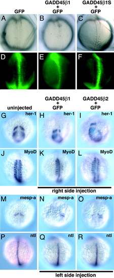
Overexpression of gadd45β genes affects somite segmentation. Synthetic RNA (gadd45β, 75 pg) together with EGFP RNA (300 pg; tracer) was injected into one blastomere of two-cell-stage embryos. The injection side was identified by EGFP before in situ hybridization. Dorsal view, anterior is up (A?F, J?L, and P?R). Posterior view, dorsal is up (G?I and M?O). (A?C) Morphology of developing somites in live embryos at 8- or 10-somite stages. (D?F) GFP expression at the corresponding embryos. Injection of zebrafish gadd45β1, but not a gadd45β1 mutant lacking middle- and C-terminal domain, caused defects in somite segmentation (A?C). (G?R) Whole-mount in situ hybridization with probes shown at the upper right. Expression of segmentation genes (mesp-a, her1, and MyoD) was severely reduced in the injected side (G?O), whereas ntl expression was not changed (P?R). EXPRESSION / LABELING:
|
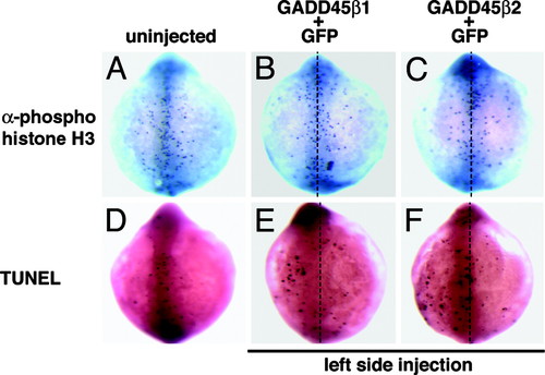
Injection of gadd45β1 RNA weakly affects cell proliferation and apoptosis. Synthetic RNA (gadd45β1, 75 pg) together with EGFP RNA (300 pg; tracer) was injected into one blastomere of two-cell-stage embryos. Dorsal view, anterior is up (A?F). (A?C) Detection of mitotic cells by immunostaining of anti-phosphohistone H3 antibody. The number of mitotic cells is slightly decreased in the injected side of gadd45β1 or gadd45β2 RNA-injected embryos. (D?F) Detection of apoptotic cells by TUNEL assay. A slightly increased number of apoptotic cells was observed at the injected side of gadd45β1 or gadd45β2 RNA-injected embryos. |
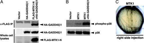
Biochemical activities of zebrafish GADD45β1. Indicated expression constructs (4 μg total: HA-GADD45β1, 2 μg; FLAG-MTK1-N, 2 μg; MTK1, 2 μg; pCS2 vector, 2 μg) were transfected into human 293T cells. Cell lysates were prepared after 24 h. (A) Cell lysates were immunoprecipitated with anti-FLAG gel and analyzed by Western blotting using anti-HA antibody (Top). Whole-cell lysates were used as controls for expression levels. (B) Phosphorylation state of p38 in the transfected cells. Cell lysates were analyzed by Western blotting using antibodies recognizing phospho-p38 and total p38. (C) Live embryos at the 10-somite stage. Synthetic RNA (human MTK1, 300 pg + EGFP, 300 pg) was injected into one blastomere of two-cell-stage embryos, leading to severe somite defects in the injection side. |
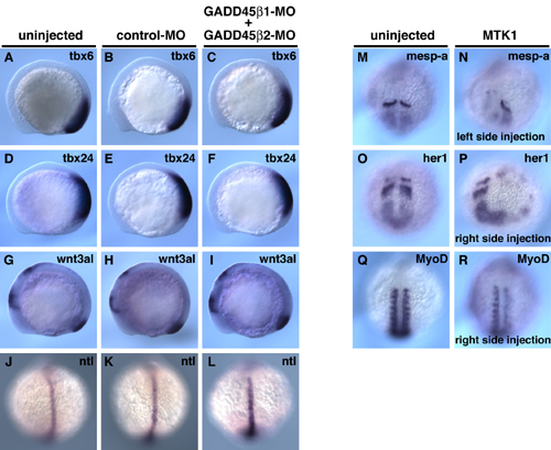
This figure illustrates that knockdown of Gadd45 expression does not affect general mesodermal markers in the embryo. The overexpression of MTK1/MEKK4 leads to reduced and disorganized expression of somite pattern genes. (A?L) Effect of gadd45β MO on marker gene expression. MOs were injected into one- or two-cell-stage embryos, as shown in Fig. 3. Lateral view, anterior is left (A?I). Dorsal view, anterior is up (J?L). Expression of tbx6, tbx24, wnt3l, and ntl was not affected in the double gadd45β-MO-injected embryos. (M?R) Effect of MTK1/MEKK4 RNA overexpression on marker gene expression. Posterior view, anterior is up (M?P). Dorsal view, anterior is up (Q and R). Expression of mesp-a, her1, and MyoD was partly suppressed, and the pattern was disorganized in MTK1 RNA-injected embryos. EXPRESSION / LABELING:
|