
- Title
-
Two distinct cell populations in the floor plate of the zebrafish are induced by different pathways
- Authors
- Odenthal, J., van Eeden, F.J., Haffter, P., Ingham, P.W., and Nüsslein-Volhard, C.
- Source
- Full text @ Dev. Biol.
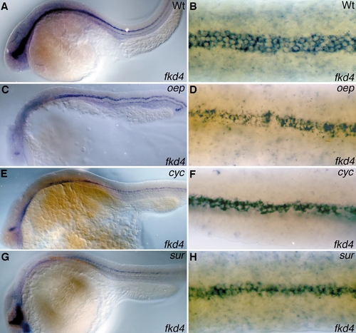
shh in the notochord can induce LFP cells. Lateral views (A, C, E, G) and dorsal views (B, D, F, H) of wild-type (A, B), oeptz257 (C, D), cyctf219 (E, F), and surty68b (G, H) embryos hybridized with probes specific for fkd4 (A–F) and fkd4/shh (G, H) in the pharyngula period (anterior is left). Only ventral neural tube cells overlying the notochord in the trunk of oep and cyc mutants form fkd4-expressing LFP cells (D, F), while in the diencephalon expression is restricted to more dorsal cells (C, E). In sur mutants shh and fkd4 expression in the forebrain appears normal (G), while fkd4 transcription in the trunk is restricted to ventral cells in contact with the notochord (H). |
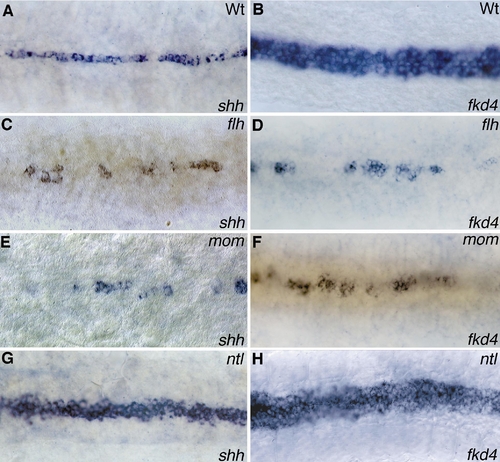
shh in the MFP can induce LFP cells. Dorsal views (anterior is left) of shh (A, C, E, G) and fkd4 (B, D, F, H) expression in wild-type (A, B), flhtk241 (C, D), momth211 (E, F), and ntltc41 (G, H) embryos in the pharyngula period. Cells around shh-expressing MFP cells in flh and mom embryos express fkd4 in the ventral neural tube (D, F). In ntl embryos cells adjacent to the broadened MFP transcribe fkd4, suggesting that in the absence of a notochord shh expression in the MFP can induce LFP development. |
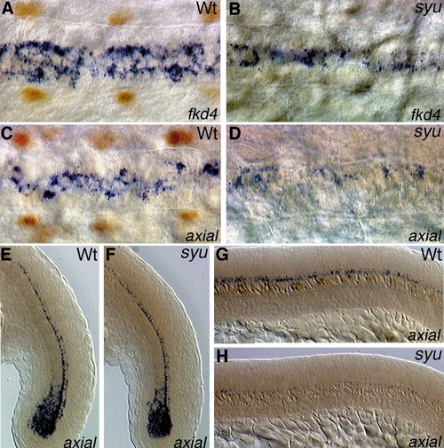
shh is required for axial and fkd4 expression in LFP cells. Dorsal views (A–D) and lateral views (E–H) of wild-type (A, C, E, G) and syut4 (B, D, F, H) embryos at the pharyngula period labeled with an in situ probe for fkd4 (A, B; blue cytoplasmic staining) or axial (C–H; blue cytoplasmic staining) and stained with the 4D9 antibody against En-like proteins (brown nuclear staining, out of focus); anterior is left. shh function is required for the induction of fkd4 and axial expression in LFP cells (B, D). While fkd4 in syu embryos stains MFP cells also in anterior trunk regions (B) axial transcription in MFP cells close to the tail bud appears normal (F), while older MFP cells maintain only low levels of expression (D, H), indicating a maintenance function of shh on axial expression in MFP cells. |
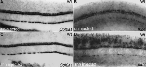
shh injection induces LFP but not MFP expression in the trunk. Lateral views of wild-type embryos in the pharyngula period stained for the MFP marker col2A1 (A, C) and the fork head domain gene fkd4 expressed in the MFP and LFP cells (B, C) after control injections (A, B) and synthetic shh RNA injections (C, D) at the one- to four-cell stage. While col2A1 expression after ectopic shh overexpression appears unaltered, fkd4 expression is dorsally expanded, indicative of a response of LFP cells to shh overexpression, whereas MFP cells appear to be nonresponsive. |
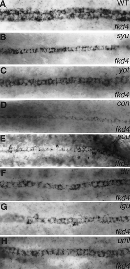
Members of the shh signal transduction cascade are involved in the induction of LFP cells. Dorsal views (anterior is left) of wild-type (A) and syutd4 (B), yotty119 (C), contf18b> (D), youty97 (E), dtrts269 (F), iguts294e (G), and umlty254z (H) mutant embryos in the pharyngula period hybridized with a probe specific for fkd4. In the ventral neural tube of the trunk region fkd4 is expressed in a three- to four-cell-broad domain including the MFP and the LFP cells. Members of the shh signal transduction cascade necessary for the induction of adaxial cells and muscle pioneer cells in the somites, syu, yot, con, and you, are also involved in the induction of LFP cells, while MFP development in these mutants appears normal. Further components of the shh signal transduction cascade are dtr (F), igu (G), and uml (H), which, similar to the you-type mutants, have defects in the projection of retinal axons, a curled-down body axis, and defects in LFP cells. While in dtr and uml embryos fkd4 staining is restricted to the MFP (F, H), in igu embryos a few fkd4-expressing cells can also be detected lateral to the MFP (G). |
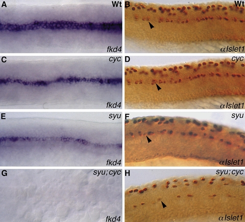
syu;cyc double mutants lack all floor plate structures in the trunk and primary motor neurons are strongly reduced. Dorsal (A, C, E, G), and lateral views (B, D, F, H; anterior is left) of wild-type (A, B), cyctf219 (C, D), syutbx392 (E, F), and syu;cyc double-mutant (G, H) embryos stained with probes specific for fkd4 (A, C, E, G) and an antibody against Islet1 (B, D, F, H) in the pharyngula period. In cyc embryos fkd4-expressing LFP cells are formed (C), whereas in syu embryos MFP cells are present and LFP are not formed (E). In syu;cyc double mutants fkd4-expressing cells in the ventral neural tube can be detected, indicating the complete absence of floor plate structures in these double mutants (G). However, fkd4 expression in the hypochord underneath the notochord and in the hindgut remains unchanged (data not shown). Primary motor neurons stained with an antibody against Islet1 are present in cyc and syu single mutants (arrowhead in D, F), but strongly reduced in syu;cyc double mutant embryos (arrowhead in H), indicating that shh together with a signal(s) from the MFP induces primary motor neurons. The few remaining motor neurons might be induced by ehh expression in the notochord. |
Reprinted from Developmental Biology, 219(2), Odenthal, J., van Eeden, F.J., Haffter, P., Ingham, P.W., and Nüsslein-Volhard, C., Two distinct cell populations in the floor plate of the zebrafish are induced by different pathways, 350-363, Copyright (2000) with permission from Elsevier. Full text @ Dev. Biol.