IMAGE

Fig. 8

ID
ZDB-IMAGE-260311-290
Genes
Source
Figures for Luo et al., 2025
Image
Figure Caption

Fig. 8

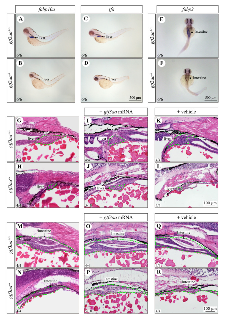
Loss of gtf3aa affects liver and intestine development. A, B In situ hybridization of fabp10a in the liver of 3-dpf gtf3aa+/+ and gtf3aa−/− larvae. C, D In situ hybridization of tfa in the liver. E, F In situ hybridization analysis of fabp2 in the intestine. G, H H&E staining of the liver in gtf3aa+/+ and gtf3aa−/− larvae. I, J H&E staining of the liver in gtf3aa+/+ larvae and gtf3aa−/− mutants rescued by gtf3aa mRNA injection at the one-cell stage. K, L H&E staining of the liver in vehicle-injected gtf3aa+/+ and gtf3aa−/− larvae. M, N H&E staining of the intestine in gtf3aa+/+ and gtf3aa−/− larvae. O, P H&E staining of the intestine in larvae rescued by mRNA injection. Q, R H&E staining of the intestine in vehicle-injected larvae. All comparisons are between gtf3aa+/+ and gtf3aa−/− groups

Figure Data
Acknowledgments
This image is the copyrighted work of the attributed author or publisher, and ZFIN has permission only to display this image to its users. Additional permissions should be obtained from the applicable author or publisher of the image. Full text @ Mar Life Sci Technol