IMAGE

Fig. 4

ID
ZDB-IMAGE-251210-49
Source
Figures for Deng et al., 2025
Image
Figure Caption

Fig. 4 
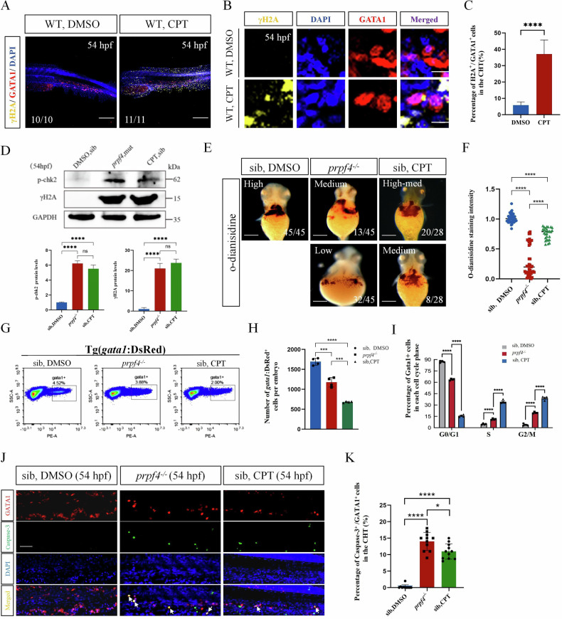
DNA damage-induced ATM/Chk2-p53 pathway partially mediates erythrocyte defects in prpf4 mutants.

A-C DNA damage examination in erythrocytes in the CHT region at 54 hpf. A Confocal images showing γH2AX (yellow) staining in WT (DMSO) and WT embryos treated with camptothecin. Scale bar: 200 µm. B High-magnification images of γH2AX and GATA1 colocalization in erythrocytes. Scale bar: 50 µm. C Quantitative analysis of γH2AX-positive cells among GATA1-positive cells in the CHT region. D Western blot analysis of p-Chk2 and γH2AX levels in 54 hpf embryos. Densitometric quantification of p-Chk2 and γH2AX protein levels is shown below. E, F Hemoglobin levels in 60 hpf embryos assessed by o-dianisidine staining. E Hemoglobin staining intensity in siblings treated with DMSO, prpf4−/− embryos, and camptothecin-treated siblings. Staining intensity is categorized as high, medium, or low. Scale bar: 100 µm. F Quantitative analysis of o-dianisidine staining intensity. G–I FACS analysis of GATA1+ erythrocytes in Tg(gata1:DsRed) embryos at 54 hpf. G The percentage of GATA1+ cells in DMSO-treated siblings, prpf4−/− embryos, and camptothecin-treated siblings (30 embryos per group). H Quantification of the total number of GATA1+ cells per embryo. I Cell cycle distribution of GATA1+ cells. Prpf4−/− embryos and camptothecin-treated siblings exhibit cell cycle arrest. J, K Detection of apoptosis in GATA1+ erythrocytes in the CHT region of 54 hpf embryos. J Confocal microscopy images showing active Caspase-3 staining in DMSO-treated siblings, prpf4−/− embryos, and camptothecin-treated siblings. Merged images highlight apoptotic signals (arrows) in GATA1+ cells. Scale bar: 50 µm. K Quantification of the percentage of active Caspase-3+ cells among GATA1+ cells in the CHT region. Both prpf4−/− embryos and camptothecin-treated siblings show a significant increase in apoptosis compared with DMSO-treated siblings.

Acknowledgments
This image is the copyrighted work of the attributed author or publisher, and ZFIN has permission only to display this image to its users. Additional permissions should be obtained from the applicable author or publisher of the image. Full text @ Cell Death Discov