
|
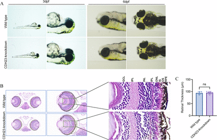
Fig. 2 (A) The ocular appearance of wild type and CDH23-knockdown zebrafish at 3 dpf and 6 dpf; (B) H&E-stained retinal sections of wild type and CDH23-knockdown zebrafish at 5 dpf; scale bars: 20 mm; (C) Effects of CDH23 knockdown on retinal thickness in zebrafish. The bar graph presents retinal thickness (μm) for wild type and CDH23-knockdown groups. Data are expressed as mean±SEM (n=5 per group); ns indicates no significant difference between groups (p>0.05, independent-samples t-test) (Representative images shown). GCL, ganglion cell layer; INL, inner nuclear layer; IPL, inner plexiform layer; IS, inner segment; ONL, outer nuclear layer; OPL, outer plexiform layer; OS, outer segment; RPE, retinal pigment epithelium.