
|
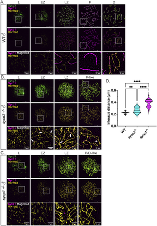
Fig. 8 Sycp1 localization to synapsed-like regions is not sufficient for Hormad1 removal. (A–C) Surface-spread chromosomes from wild-type (A), syce2-/- (B) and sycp1-/- (C) spermatocytes immunostained for Sycp3 (green), Sycp1 (magenta) and Hormad1 (yellow). Examples of spread chromosomes from meiotic prophase I described in Fig 7A–C. Scale bar = 10 µm. Boxes represent magnified areas for each stage. Scale bar for magnified examples = 2 µm. Arrows represent Hormad1 localization to pseudo-synapsed regions with Sycp1. (D) Violin plot showing the distance between axes for co-aligned axial pairs in early zygotene for wild-type (n = 28), syce2-/- (n = 28) and sycp1-/- (n = 28) spermatocytes. 4 cells from each genotype were used for the analysis. Significance was determined using ordinary one-way ANOVA testing with uncorrected Fisher’s LSD test. ** = p < 0.01; *** = p < 0.001; **** = p < 0.0001.