
|
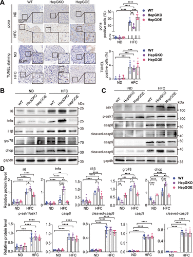
Fig. 5 Effect of hep-gmfb abundance on the molecular functional pathways in MASLD liver. A: representative anti-pcna immunochemical images and TUNEL staining images in liver sections from ND and HFC groups of WT, HepGKO, and HepGOE zebrafish, and quantification of the positive staining cells. Scale bar = 50 μm. n = 8 fish per group. One-way ANOVA, **P < 0.01, ***P < 0.001, ****P < 0.0001 vs. ND or WT group. B–D: expression levels of proteins il6, tnfα, il1β, grp78, and chop (B); and ask1, p-ask1, casp8, cleaved-casp8, casp9, and cleaved-casp9 (C) with gapdh as normalization control in liver samples from ND and HFC groups of WT, HepGKO, and HepGOE zebrafish. n = 6 samples for liver tissue, 10 fish per sample. Data were presented as relative expression levels (D). One-way ANOVA, *P < 0.05, **P < 0.01, ***P < 0.001, ****P < 0.0001 vs. ND or WT group. Experiments were repeated on at least 3 clutches. ask1, apoptosis signal regulating kinase-1; casp 8, caspase 8; casp 9, caspase 9; chop, C/EBP homologous protein; gapdh, glyceraldehyde-3-phosphate dehydrogenase; grp78, glucose-regulated protein 78; hep-gmfb, gmfb in hepatocyte; HepGKO, hepatocyte-specific gmfb knockout; HepGOE, hepatocyte-specific gmfb overexpression; HFC, high fat, high cholesterol diet; il1β, interleukin 1 beta; il6, interleukin 6; MASLD, metabolism-associated steatotic liver disease; ND, normal diet; pcna, proliferating cell nuclear antigen; tnfα, tumor necrosis factor alpha; TUNEL, terminal dexynucleotidyl transferase (TdT)-mediated dUTP nick end labeling; WT, wild-type.