
|
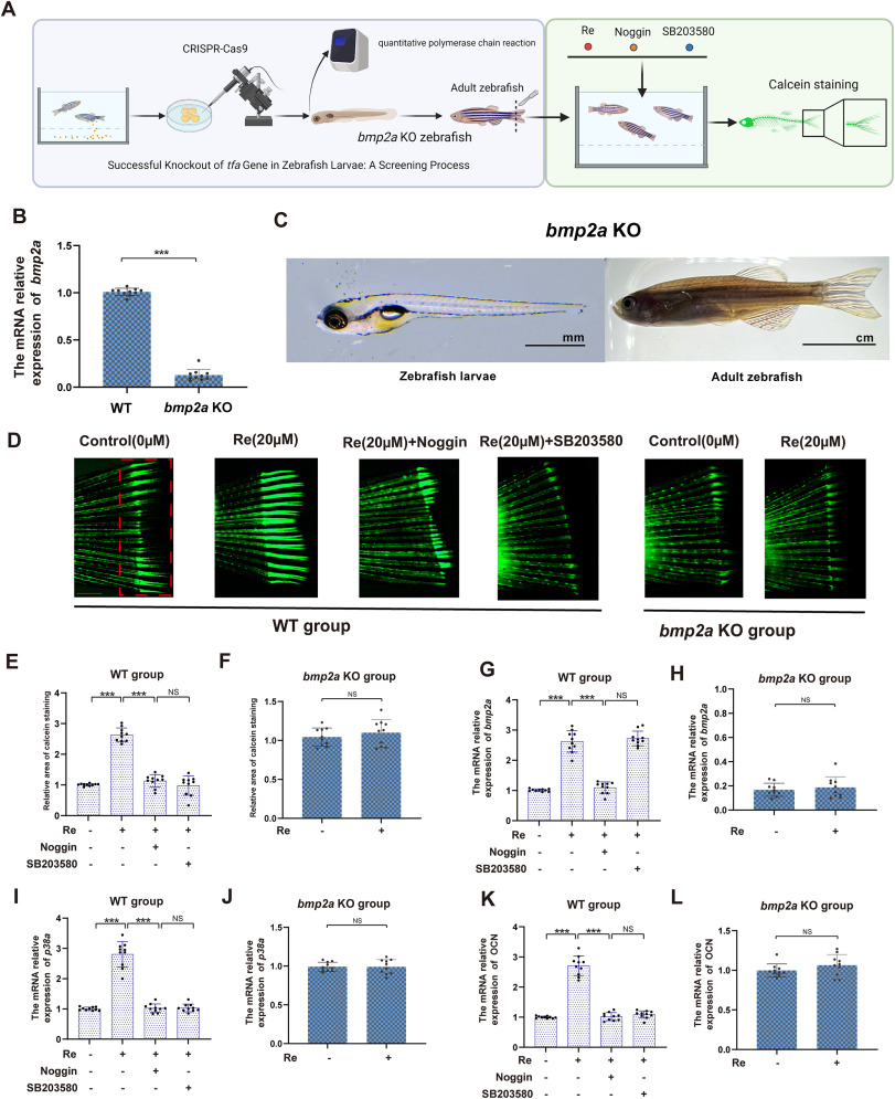
Fig. 6 Ginsenoside Re modulates bone formation via the Bmp2/p38 pathway in vivo. A. The in vivo experimental demonstration illustrates the construction of the bmp2a knockout zebrafish model. After tail amputation surgery, both the bmp2a KO group and the control group were co-cultured with Re, noggin, and SB203580 for 7 days. The effects of Re, bmp2a and the p38 pathway(mapk14a) on zebrafish bone formation were evaluated using calcein green staining. B-C. After constructing the bmp2a knockout zebrafish model, qPCR technology was utilized to verify the knockout results. Additionally, the appearance of zebrafish larvae and adult fish after bmp2a knockout was demonstrated. D-F. The bone formation in the tail fin of the zebrafish models from the control group and the bmp2a KO group was assessed using calcein staining after treatment with Re, noggin and SB203580. G-L. The relative expression levels of osteocalcin, bmp2a, mapk14a mRNA in the tail fin of the zebrafish models from the bmp2a knockout group and the control group after treatment with Re, noggin and SB203580. Statistical methods: two-tailed Student's t-test, (∗P < 0.05, ∗∗P < 0.01, ∗∗∗P < 0.001).