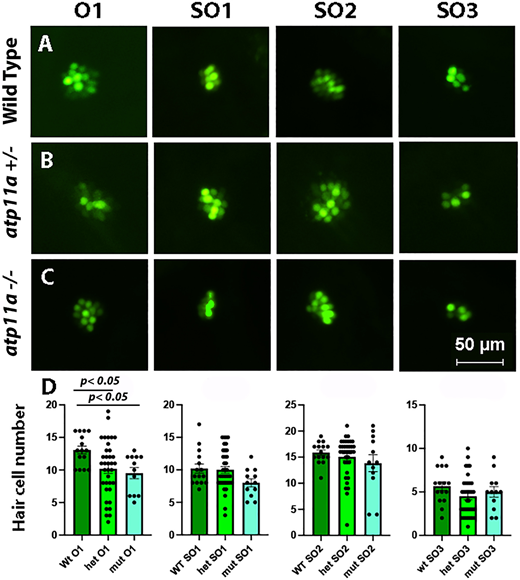
Fig. 3 Reduced number of hair cells in the O1 neuromast. (A–C) Nuclear staining of four neuromasts in 5 dpf atp11anl1007 larvae with Yo-Pro-1, demonstrating a decrease in the number of positively stained cells in the O1 neuromast in homozygous and heterozygous mutants compared with wild type. (D) Neuromast hair cell quantification for one otic (O1) and three supraoptic (SO1, SO2 and SO3) neuromasts demonstrates a significant reduction in hair cell number for the O1 neuromast in homozygous and heterozygous mutants. Data are mean±s.e.m. with significance calculated using a one-factor ANOVA with Tukey's post-hoc analysis for multiple comparisons. Wild type, n=15; heterozygous, n=36; homozygous mutant, n=12.
                
                    
                        Image


                        Figure Caption


                        Figure Data


                        Acknowledgments


This image is the copyrighted work of the attributed author or publisher, and
ZFIN has permission only to display this image to its users.
Additional permissions should be obtained from the applicable author or publisher of the image.
      Full text @ J. Cell Sci.