
Fig. 5
- ID
- ZDB-IMAGE-250602-25
- Publication
- Tian et al., 2025 - C-mannosyltransferase DPY19L1L-mediated Reissner Fiber formation is critical for zebrafish (Danio rerio) body axis straightening
- All Figures
- Figures for Tian et al., 2025
|
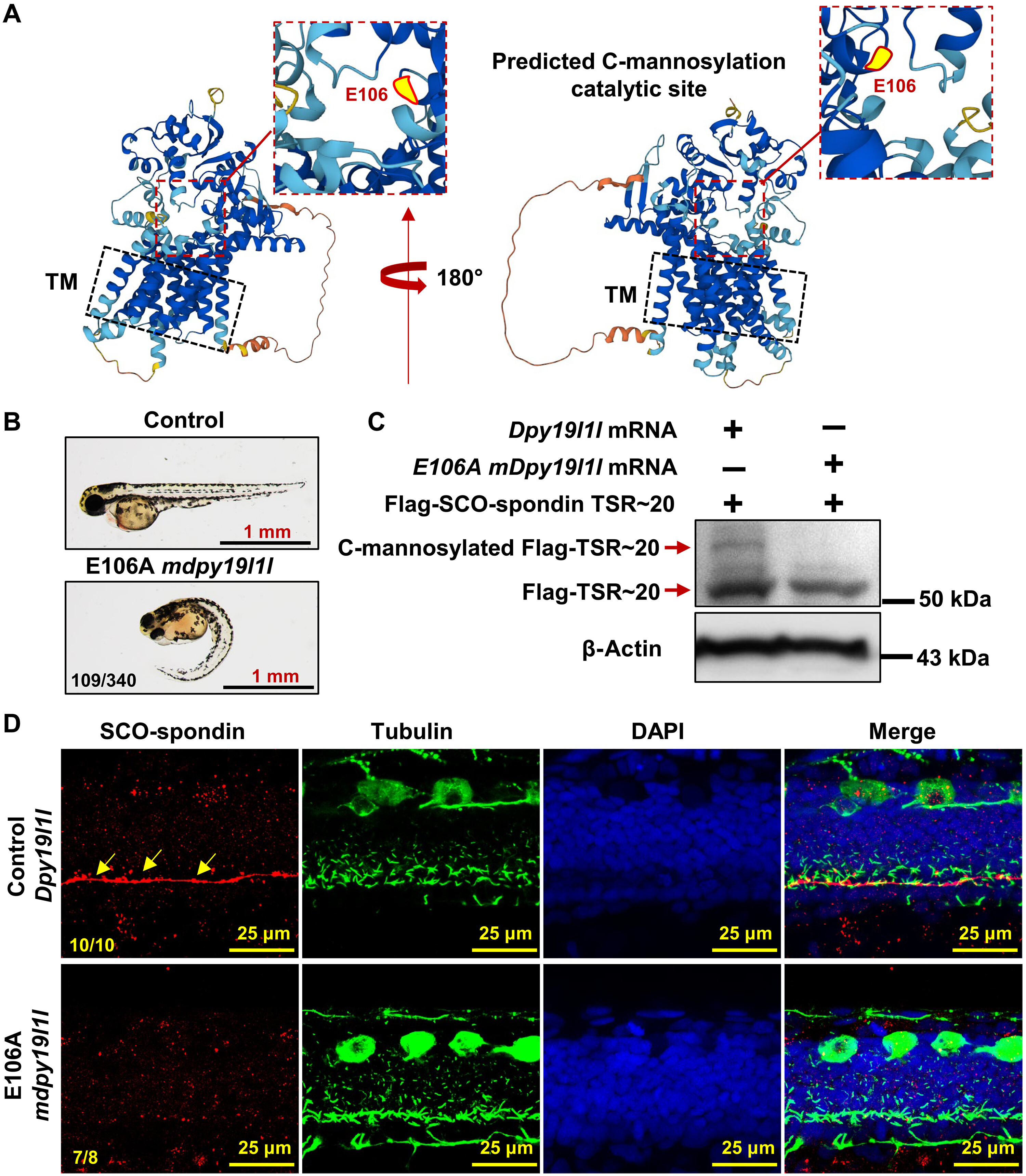
Fig. 5 DPY19L1L C-mannosylates SCO-spondin TSR motifs. (A) AlphaFold-predicted DPY19L1L structure. Red box highlights the catalytic core; yellow dot marks putative C-mannosylation site (E106). TM, transmembrane region. (B) Phenotypes of wild-type embryos injected with E106A dpy19l1l mRNA at 48 hpf. (C) Immunoblot of Flag-TSR~20 lysates from injected embryos. Anti-Flag detects Flag-TSR~20 and C-mannosylated Flag-TSR~20. (D) RF localization (red) in wild-type embryos injected with control or E106A dpy19l1l mRNA (36 hpf). Red: SCO-spondin; green: cilia; DAPI: nuclei. Yellow arrows indicate RF in controls.