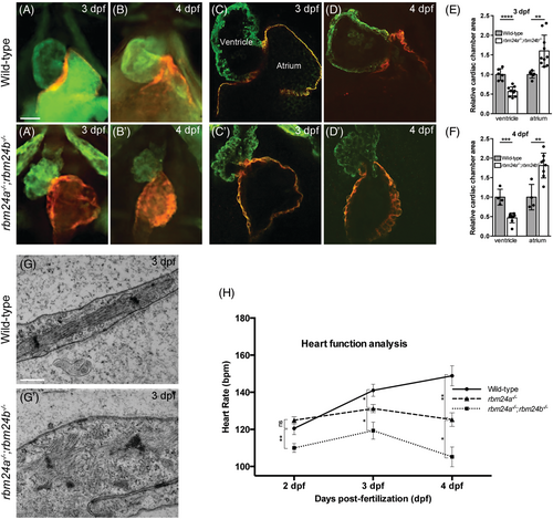
Fig. 7 Disrupted integrity and function of cardiac muscles in rbm24a and rbm24b mutants. (A–D′) Immunofluorescence staining of ventricle and atrium in wild-type embryos and rbm24a−/−;rbm24b−/− double mutants. At each developmental stage, 15 to 20 embryos per genotype were analyzed. Scale bars: 5 μm. (E, F) Graphs measuring the ventricle and atrium size at 3 and 4 dpf, respectively. (G, G′) TEM images of heart sections. Wild-type embryos show highly organized sarcomeres with thin and thick myofilaments in well-aligned bundles and discernible dark A-band and light I-bands, while double mutant embryos display a complete disorganization of sarcomeric unit. At each developmental stage, 15 to 20 embryos per genotype were analyzed. Scale bars: 500 nm. (H) Graph comparing heart rate of rbm24a single mutants and rbm24a−/−;rbm24b−/− double mutants at 2, 3, and 4 dpf. Average heart rate with standard error was plotted and significant deviation was determined using the Student's t test. At each developmental stage, 15 to 20 embryos per genotype were analyzed.
Image
Figure Caption
Figure Data
Acknowledgments
This image is the copyrighted work of the attributed author or publisher, and
ZFIN has permission only to display this image to its users.
Additional permissions should be obtained from the applicable author or publisher of the image.
Full text @ Dev. Dyn.

