
Image
|
Figure Caption
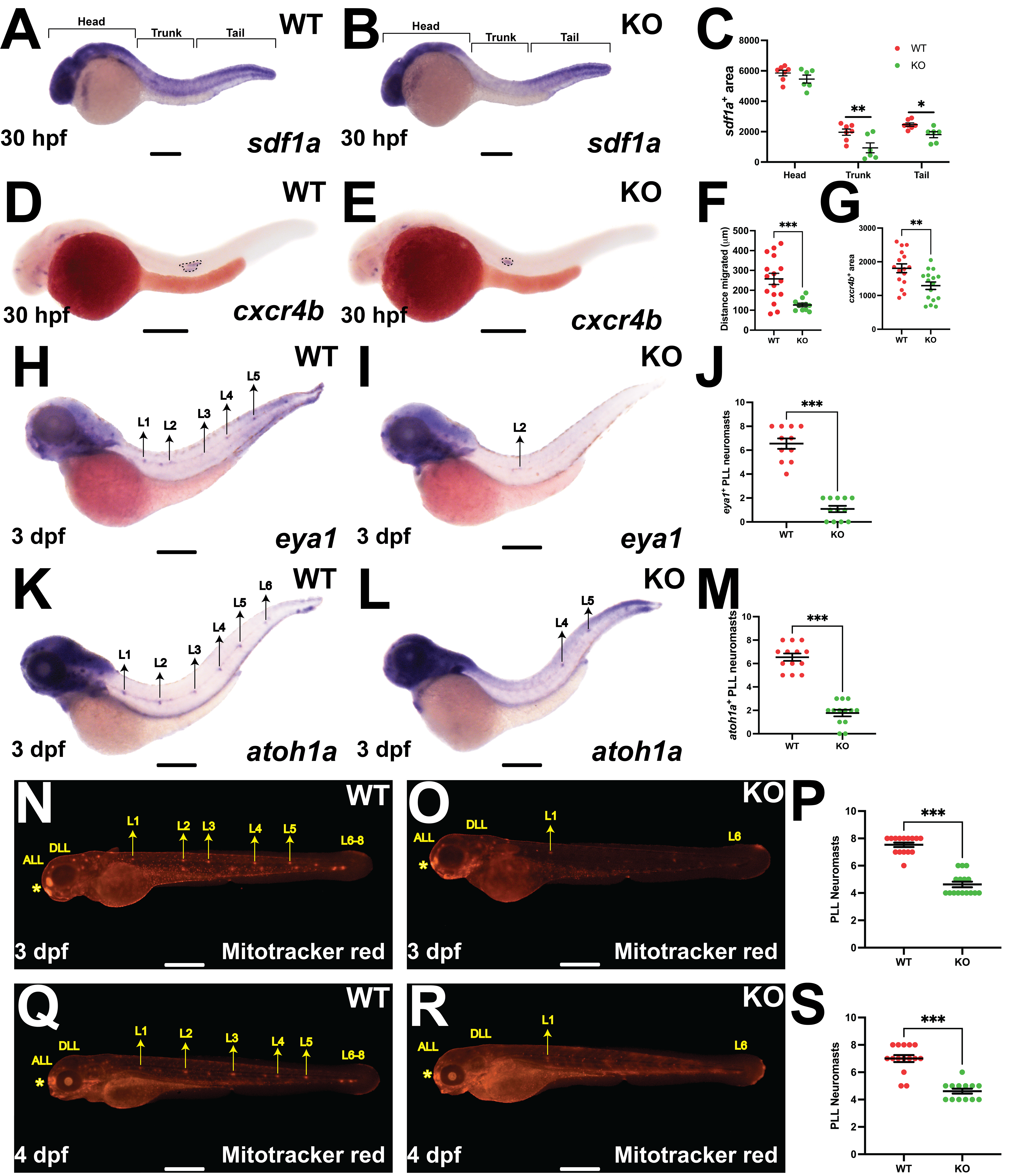
Fig. 5 Analysis of potential pathway components. (A–C) Representative images of wildtype (WT), Socs3b knockout (Socs3b KO) and Stat3 knockout (Stat3 KO) embryos analyzed with Mitotracker red at 3 dpf, with scale bars of 100 µm shown. (D) Quantitation of PLL neuromasts, showing individual points along with the mean ± SEM. No statistically significant differences were observed between groups (n = 14–16, repeated twice). (E) RT-PCR analysis of the indicated genes in WT, Socs3a KO and Socs3b KO embryos at 3 dpf
Acknowledgments
This image is the copyrighted work of the attributed author or publisher, and
ZFIN has permission only to display this image to its users.
Additional permissions should be obtained from the applicable author or publisher of the image.
Full text @ Front Biosci (Landmark Ed)