
|
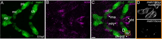
Fig. 2 - Supplement 2 matn1 is expressed in differentiating cranial tenocytes. Ventral view of the developing mandibular arch in a 51 hpf embryo showing in situ Hybridization Chain Reaction (isHCR) of matn1 (A, C, D) and scxa (B–D). (D) magnified view of yellow ROI (C) shows outline of tenocyte nuclear 3D volume with white puncta representing voxel colocalizations of matn1 and scxa as depicted by colocalization using Imaris (see methods). ep = ethmoid plate cartilage, ch = ceratohyal cartilage, mc = meckel’s cartilage, pq = palatoquadrate cartilage, ch-pqt = ceratohyal-palatoquadrate tendon, ima = intermandibularis anterior tendon, mhj = mandibulohyoid junction tendon, sht = sternohyoideus tendon. Scale bars = 20 µm.