
|
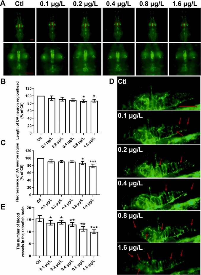
Fig. 2 UV-531 induces DA neuron damage, cerebral vessels deficits in zebrafish. (A) Fluorescence microscopy images of slc18a2:GFP zebrafish exposed to UV-531. Red brackets indicate DA neurons. Images were enlarged to improve visualization of DA neuron morphology. Scale bar, 150 μm. n = 15. (B) Statistical analysis of the length of DA neuron/head in each group (% of Ctl). n = 15. (C) Statistical analysis of the fluorescence of DA neuron region (% of Ctl) in each group. n = 15. (D) Representative fluorescence microscopy images of fli1:GFP zebrafish exposed to UV-531 up to 4 dpf. Red arrows indicate vessel damage. Scale bar, 150 μm. n = 15. (E) Statistical analysis of the number of blood vessels in the brain. n = 15. *p < 0.05, * *p < 0.01, * **p < 0.001 vs. Ctl.