
Fig. 3
- ID
- ZDB-IMAGE-250430-13
- Genes
- Publication
- Fragale et al., 2025 - Loss of Atoh8 Affects Neurocranial and Axial Skeleton Development in Zebrafish
- All Figures
- Figures for Fragale et al., 2025
|
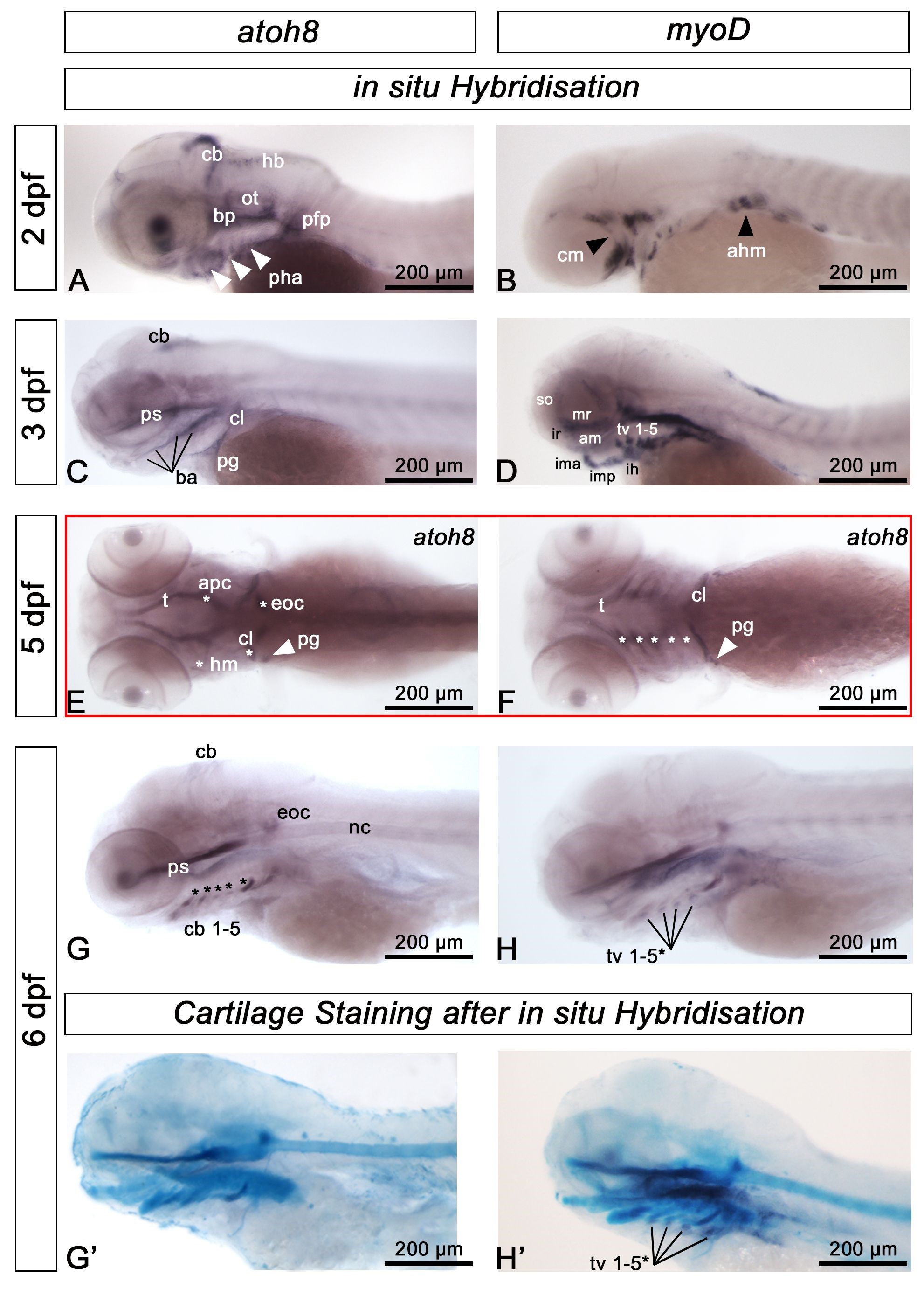
Fig. 3 atoh8 expression in larval skeletal elements.In situ mRNA hybridisation for atoh8 and myoD mRNA in wild type zebrafish at the indicated late embryonic and early larval stages. (A,B) At 2 dpf, the expression pattern is distinctly different for each gene. atoh8 mRNA is largely expressed in precursor cells of the cartilage anlagen including the branchial arches (white arrowheads) and in the developing cerebellum (cb), hindbrain (hb) and pectoral fin primordium (pfp), whereas myoD mRNA labels undifferentiated myogenic precursors of cranial (white arrowhead, cm) and anterior hypaxial muscle (black arrowhead, ahm). (C,D) atoh8 mRNA is detected in the branchial arches and marks craniofacial skeletal anlagen, whereas myoD mRNA persists in skeletal muscle precursors. (E,F) atoh8 mRNA in craniofacial elements; dorsally: trabecula (t), anterior parachordal (apc), hyomandibular (hm), cleithrum (cl) and the pectoral girdle (arrowhead, pg), ventrally: trabecula (t), cleithrum (cl), pectoral girdle, and ceratobranchials (cb) 1-5 (asterisks). (G) atoh8 mRNA in derivatives of the branchial arches, labelling a population of cells associated with ceratobranchial cartilages 1-5 (arrowheads). (H) myoD expression goes on in myogenic precursor cells. (G’,H’) atoh8 and myoD expression, respectively, on Alcian Blue stained larvae, showing that atoh8 and myoD labelled cells associated with the ceratobranchials occupying different locations; myoD in (H’) labels the transversus ventralis (tv) muscles 1-5; *the most anteriorly located tv 1 is not labelled. All except (E,F): anterior to the left, dorsal up; (E) dorsal view, (F): ventral view. Scale bars: 200 µm.