
|
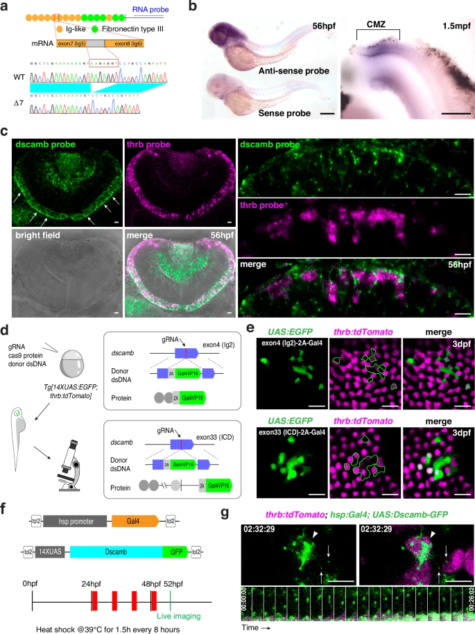
Fig. 2 Dscamb is localized in apical filopodium-like neurites of cone photoreceptors.a Zebrafish Dscamb protein has conserved Ig-like and FNIII domains in the extracellular domain. In addition to one full-length isoform, we detected cDNA encoding a 7-bp-deletion isoform. An RNA probe is designed in the intracellular domain. b In situ hybridization of 56-hpf zebrafish embryos (left) and 1.5-mpf dissected retinas (right) with a dscamb RNA probe. dscamb mRNA is expressed in differentiating photoreceptors near the retinal ciliary marginal zone (CMZ). Three independent experiments. c In situ hybridization of 56-hpf retinas with dscamb (green) and thrb (magenta) RNA probes. dscamb mRNA is expressed in photoreceptor layer (white arrows in left upper panel). Higher magnification images (right panels) indicate that dscamb mRNA signals were detected in both thrb-positive (red cones) and -negative photoreceptors. Three independent experiments. d CRISPR/Cas9-mediated strategy of in-frame knock-in of 2A-Gal4VP16 DNA into exon 4 and exon 33 of the dscamb gene. e Mosaic generation of photoreceptors in which 2A-Gal4VP16 DNA was inserted in-frame in exon 4 (upper panels) and exon 33 (bottom panels) of the dscamb gene in 3-dpf Tg[UAS:EGFP; thrb:tdTomato] transgenic retinas. tdTomato labels red cones. EGFP (green) was expressed in both red cones and non-red cone photoreceptors in both cases. Three independent experiments for each exon trapping. f Experimental design to visualize GFP-tagged Dscamb in cones. Transgenic embryos carrying Tg[hsp:Gal4; UAS:Dscamb-GFP] were used for live scanning at 52 hpf after multiple heat-shock treatments. g Live imaging of apical surfaces of red cones in transgenic embryos carrying Tg[hsp:Gal4; UAS:Dscamb-GFP; thrb:tdTomato] at 52 hpf. tdTomato labels red cones. GFP-tagged Dscamb was accumulated on apical domains (arrowheads) and at tips of filopodia (arrows). The time series montage (bottom panels) indicates that GFP-tagged Dscamb localization at the filopodial tip is maintained during filopodial extending/retracting cycle. Scale bars: 300 μm b, 10 μm c, e and 5 μm g