
|
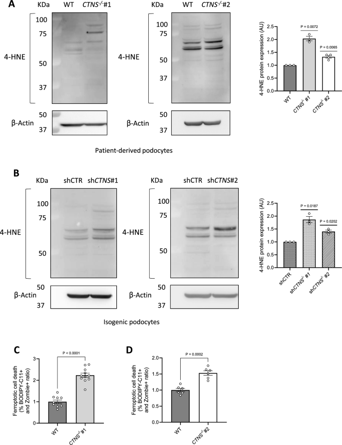
Fig. 4 Cystinosis podocytes present increased 4-hydroxynonenal (4-HNE) protein expression and ferroptotic cell death. (a, b) Representative western blot analysis of 4-hydroxynonenal (4-HNE)-conjugated protein levels. a. Left panel: WT and CTNS−/−#1; central panel: WT and CTNS−/−#2, with β-Actin used as the loading control. Right panel: quantification of 4-HNE protein expression normalized to β-Actin. (n ≥ 3 biological experiments, n = 1 technical replicate). Statistical analysis: one-sample t-test, with WT set as the reference. b. Left panel: shCTR and shCTNS#1; central panel: shCTR and shCTNS#2, with β-Actin used as loading control. Right panel: quantification of 4-HNE protein expression normalized to β-Actin. (n = 3 biological experiments, n = 1 technical replicate). Statistical analysis: one-sample t-test, with shCTR set as the reference. (c, d) Ratio of the percentages of BODIPY-C11 + cells among dead podocytes (Zombie) measured by flow cytometry. c. CTNS−/−#1 normalized to WT; d. CTNS−/−#2 normalized to WT. (n ≥ 3 biological experiments, n = 1 technical replicate). Statistical analysis: Welch’s unpaired t-test Each dot represents a biological experiment