
|
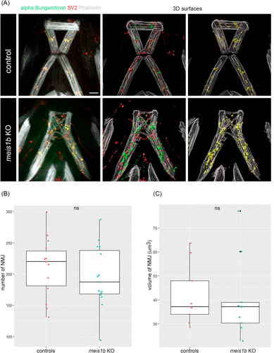
Fig. 10 Neuromuscular junctions (NMJ) are developed and colocalized in meis1b mutants. (A) Left panels: Combined antibody staining of presynaptic receptors (SV2, red) and postsynaptic receptors (alpha bungarotoxin, green). Muscles are counterstained with phalloidin (white). Middle and right panels: 3D surfaces of images on the left generated in Imaris (n = 15), yellow indicates colocalization of SV2 and alpha bungarotoxin (NMJ). (B) Quantification of the number of NMJ, (C) volume of NMJ. Color dots indicate individual animals (controls = 9, mutants = 9). Two tailed t-test was used for statistical analysis. All images are ventral views, anterior on top. Scale bar = 30 μm.