
Image
|
Figure Caption
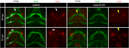
Fig. 8 Expression of scxa in tenocytes surrounds the tips of the Meckel's cartilage. HCR-antibody staining of scxa mRNA (red) and cartilage (Sox9 antibody, green). Ventral views with the anterior to top. In controls at 60–72 hpf, the anterior tenocyte cluster in the midline was localized between and anterior to the tip of the Meckel's cartilage (arrows). In contrast, this scxa-positive cluster was missing in meis1b mutants (arrowheads). ch, ceratohyals; mc, Meckel's cartilage; pq, palatoquadrate. Scale bar = 30 μm.
Acknowledgments
This image is the copyrighted work of the attributed author or publisher, and
ZFIN has permission only to display this image to its users.
Additional permissions should be obtained from the applicable author or publisher of the image.
Full text @ Dev. Dyn.