
|
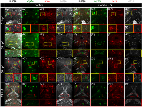
Fig. 7 Tenocytes development is affected together with muscles in meis1b mutants. Tenocytes were analyzed by HCR FISH using probes for xirp2a (green, A′–D′, E′–H′) and scxa (red, A″–D″, E″–H″) and muscles were co-stained by MF20 antibody (white, A‴–D‴, E‴–H‴). Ventral views with the anterior to the top. (A, A″) scxa expression was localized in two lateral cluster (yellow dotted frame and insets) and one in the middle (red dotted frame and insets). In meis1b mutants (E–E″), expression of scxa is seen only in two lateral clusters (yellow dotted frame and insets) whereas the expression in the middle is missing (red dotted frame and insets). (A–A‴, E–E‴) Detail of the tip of adductor mandibulae in yellow insets, detail of the anterior scxa positive area in red insets. (B′, F′) xirp2a is expressed at the muscle tips in the presumptive mandibulohyoid junction (yellow dotted frame and insets). (B–B‴, F–F‴) Detail of the mandibulohyoid junction in yellow insets. (C, G) xirp2a and scxa are co-expressed at the sides of attachment to the Meckel's cartilage (yellow dotted frame and insets). In meis1b mutants (G′), xirp2a expression is less condensed in the mandibulohyoid junction (red dotted frame and insets). (C–C‴, G–G‴) Detail of the presumptive intermandibular tendon in yellow insets, detail of the mandibulohyoid junction in red insets. (H) Loss of the hyohyal muscle led to an underdeveloped the hyohyal junction (red dotted frame and insets). (D–D‴, H–H‴) Detail of the hyohyal junction in red insets. Scale bar = 30 μm.