
|
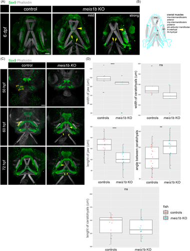
Fig. 4 Muscle fiber structure is modulated by impaired cartilage in meis1b mutants. Antibody staining of cartilage (Sox9, green) and muscles (phalloidin, white). Ventral views, anterior to the top. (A) At 6 dpf, separated muscle fibers of the intermadibularis posterior (imp) are indicated by yellow arrowheads. Split ends of the hyohyal muscles in mutants are labeled with yellow arrows. (B) Schematic of cranial muscles (gray) and contours of adjacent cartilage (blue). (C) At stage 50 hpf, rudiments of the Meckel's cartilage (mc), palatoquadrate (pq) and ceratohyals (ch) start to form, the only visible muscle is the adductor mandibulae (am) at this stage. At 60 hpf, the separated intermadibularis posterior muscle fibers are already detected at this stage (arrowhead). The hyohyal muscle are underdeveloped (arrows). At 72 hpf, the intermadibularis posterior muscle fibers are unaligned (arrowhead) and the fibers of the hyohyal muscle appear separated even more (arrows). Scale bar = 30 μm. (D) Quantification of the width of the jaw and ceratohyals at 50 hpf (controls = 5, mutants = 5), length of the jaw and ceratohyals and angle between the ceratohyals at 72 hpf (controls = 16, mutants = 16). Two tailed t-test was used for statistical analysis, **p < .01, ***p < .001. am, adductor mandibulae; ch, ceratohyals; mc, Meckel's cartilage; pq, palatoquadrate. Scale bar = 50 μm.