
|
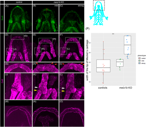
Fig. 3 Chondrocytes of the Meckel's cartilage appear unaligned in meis1b KO mutants. (A–C) Whole-mount antibody staining of chondrocytes with Sox9 (green) and (D–O) Col2 (magenta) at 8 dpf. (A–I), (M–O), Ventral views; (J–L), lateral view; anterior to the top. The position of cartilage in images is displayed in a dotted frame in the scheme (right). (G–I) Magnification of dotted frame of (D–F), the border between Meckel's cartilages is missing in meis1b mutants (H, I). (H, I) Chondrocytes are misaligned which causes irregularities in the cartilage (red arrowheads). (J–L) Magnification of dotted frame of (D–F) from lateral view; The posterior end of the Meckel's cartilage (white arrows) and anterior end of palatoquadrate (red arrows) are deformed. The gap within the jaw joint is missing. (M–O) Optical section of the Meckel's cartilage from (D–F). (M) Chondrocytes are stacked in columns of columnar-shaped cells. (N, O) The shape of chondrocytes is uneven and chondrocytes are unorganized. The red line indicates the site of width measurement of Meckel's cartilage. (P) Quantification of the width of the tip of Meckel's cartilage. Two tailed t-test was used for statistical analysis, control (n = 17), mutants (n = 17: mild = 6, strong = 11), **p < .01. Scale bar = 30 μm.