
|
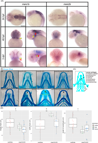
Fig. 2 Meis1b affects the jaw and ceratohyal cartilages development. (A) Expression of meis1b and meis2b in wild type embryos during formation of NC derivatives. Meis1b is expressed in mandibular (yellow arrow) and hyoid arches (red arrow). Dorsal view (24 hpf, left column), ventral view (48 hpf, 3 dpf, left column), lateral view (right column in meis1b and meis2b). Scale bar = 100 μm. (B) Staining of cartilage with Alcian Blue at stage 8 dpf. Ventral views, anterior to the top. Meis1b and meis1ab mutants were separated into two groups by the level of morphological damages: mild (n = 8) and strong (n = 24). The yellow lines indicate distance measurements. The fusion of the Meckel's cartilage is shown by arrowheads. (C) Schematic drawing of cartilage at 8 dpf, the dotted frame corresponds to images in (B). D, Quantification of the jaw length (a), width (b) and the length of the ceratohyals (c). Two tailed t-test was used for statistical analysis, controls (n = 15), mutants (n = 15: mild = 4, strong = 11), *p < .05, **p < .01. Scale bar = 50 μm.