
|
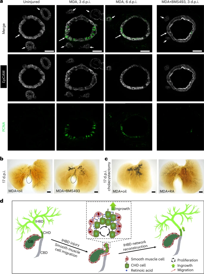
Fig. 6 The roles of the RA signal in CHD cell proliferation and IHBD network reconstruction are conserved in mouse.a, EpCAM and PCNA immunohistochemistry on sections of the CHD from uninjured mice (n = 5/5), MDA-injured mice at 3 d.p.i. (n = 4/5) and 6 d.p.i. (n = 4/5), and MDA-injured mice after BMS493 treatment at 3 d.p.i. (n = 4/6). Arrows and arrowheads indicate CHD and peribiliary glands, respectively. b,c, The IHBD system visualized by retrograde ink injection into the CBD after MDA treatment with oil (n = 9/13) and BMS493 (n = 10/12) (b) and with cholecystectomy plus MDA treatment with oil (n = 11/15) and RA (n = 11/14) (c) at 17 d.p.i. Dashed lines outline the gallbladder. d, Diagram summarizing the role of the gallbladder in reconstruction of the IHBD network after severe damage. In this process, gallbladder-derived smooth muscles coated with CHD produce RA to activate the proliferation and ingrowth of CHD cells into the inner liver, thereby reconstructing the IHBD system. Scale bars, 50 μm (a) and 200 μm (b,c).