
|
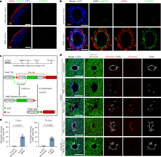
Fig. 4 Smooth muscle migration and CHD ingrowth occur after severe IHBD damage in mouse.a, αSMA and ALDH1A2 immunofluorescent confocal images of gallbladder at 4 d.p.i. in MDA-treated (n = 7/7) and uninjured (n = 7/7) mice. b, αSMA and ALDH1A2 immunofluorescent confocal images of the CHD at 4 d.p.i. in MDA-treated (n = 5/7) and uninjured (n = 5/5) mice. Note that the CHD cells are coated with αSMA and ALDH1A2 double-positive cells after MDA treatment. c, Experimental strategies for lineage tracing of EHBD and IHBD cells with Cre/loxP-DreER/rox systems. In Krt19:DreER × Alb:Cre × DeaTL-IR triple transgenic mice, EHBD cells are marked with tdTomato after 4-OHT treatment, and IHBD cells and hepatocytes are marked with ZsGreen with or without 4-OHT treatment. d, CK19, tdTomato and ZsGreen immunofluorescent confocal images of the IHBD in 4-OHT-treated only, 4-OHT- plus MDA-treated and corn oil- plus MDA-treated mice. e, Quantification of the ratio of tdTomato+ among CK19+ cells in 4-OHT-treated only and 4-OHT plus MDA-treated mice at 7 d.p.i. (n = 6 mice) and 15 d.p.i. (4-OHT plus uninjured, n = 5 mice; 4-OHT plus MDA, n = 10 mice). Data are presented as mean ± s.e.m.; unpaired t-test. Scale bars, 100 μm (a) and 50 μm (b,d).