IMAGE

Fig. 4

ID
ZDB-IMAGE-250212-61
Source
Figures for Liu et al., 2025
Image
Figure Caption

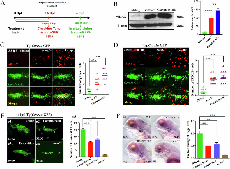
Fig. 4 Immature T cells in mcm5 mutants are more sensitive to DNA damage.

A Schematic for chemical treatment and subsequent examination. B γH2AX protein was evaluated by Western blotting. The right columns show the relative gray level. C γ-H2AX is a phosphorylated form of H2AX, which is a DNA damage marker. DNA damage was evaluated by γH2AX immunostaining for siblings (n = 10), mcm5−/− embryos (n = 10) and siblings treated with camptothecin (n = 10). D Proapoptotic Coro1a:GFP cells were examined using TUNEL staining for embryos with different treatments at 3.5 dpf. More TUNEL-positive Coro1a:GFP cells were observed in embryos treated with camptothecin. E On 4 dpf, the expression of Coro1a:GFP in the thymus was compared among siblings (e1, n = 42), embryos treated with camptothecin (e2, n = 35) or roscovitine (e3, n = 39), and mcm5−/− embryos (e4, n = 24). The number of Coro1a:GFP labeled cells was decreased in embryos treated with camptothecin or roscovitine, but the phenotype was not as strong as that in mcm5−/− embryos (e5). F Expression of rag1 in embryos with different treatments. Embryos treated with camptothecin (45% of WT controls) or roscovitine (56% of WT controls) displayed decreased expression of rag1, but the decrease was not as strong as that in mcm5−/− embryos (10.7% of controls). For (BF), the data were presented as means ± SD; The P-values (t-test; two-tailed); NS, not significant. “**” P < 0.01, “***” P < 0.001.

Acknowledgments
This image is the copyrighted work of the attributed author or publisher, and ZFIN has permission only to display this image to its users. Additional permissions should be obtained from the applicable author or publisher of the image. Full text @ Cell Death Dis.