IMAGE

Fig. 3.

ID
ZDB-IMAGE-250122-54
Source
Figures for Willoughby et al., 2024
Image
Figure Caption

Fig. 3.

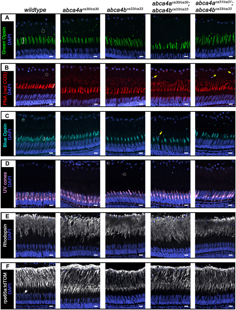
Morphology of COS, ROS and RPE in retinas of 2-month-old zebrafish. (A-F) Confocal z-projections of COS, ROS and RPE in retinas obtained from wild-type or abca4aca30/ca30, abca4bca33/ca33, abca4aca30/ca30;abca4bca33/ca33 and abca4aca31/ca31;abca4bca33/ca33 mutant zebrafish. (A) Green COS stained for Green Opsin (green; z=3 µm), (B) red COS stained for PNA (red; z=3 µm), (C) blue COS stained for Blue Opsin (cyan; z=3.4 µm), (D) UV cones expressing EGFP stained for GFP (violet; z=4.4 µm), (E) ROS stained for rhodopsin (white; z=1.9 µm) and (F) RPE expressing tdTomato stained for RFP (white, z=2.4 µm). Counterstaining for nuclei is shown in dark blue (DAPI). White arrow in F (wild-type panel) indicates distal RPE processes around UV COS (not visible). Encircled areas show examples of phagosomes in retinas of wild-type and abca4bca33/ca33 fish. White brackets in wild-type panels A-D indicate COS. Yellow arrows indicate examples of non RPE (‘mystery’) nuclei in B and C. Scale bars: 10 µm.

Acknowledgments
This image is the copyrighted work of the attributed author or publisher, and ZFIN has permission only to display this image to its users. Additional permissions should be obtained from the applicable author or publisher of the image. Full text @ Dis. Model. Mech.