
|
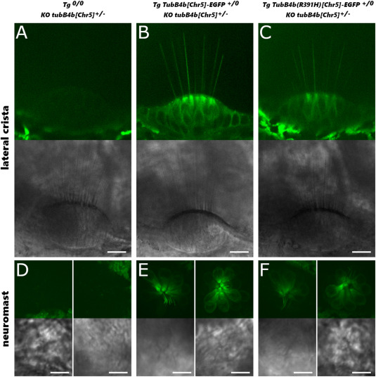
Fig. 6 Expression of TubB4b[Chr5]-EGFP and TubB4b(R391H)[Chr5]-EGFP in inner ear lateral cristae and lateral line neuromast of 5 dpf larva. KO tubB4b[Chr5]+/− larva (A,D), Tg(tubB4b[Chr5]:TubB4b[Chr5]-EGFP)+/0; KO tubB4b[Chr5]+/− larva (B,E) and Tg(tubB4b[Chr5]:TubB4b(R391H)[Chr5]-EGFP)+/0; KO tubB4b[Chr5]+/− larva (C,F) were imaged live by spinning microscopy. Single digital section in central area of cristae (A–C); maximum projection of image stacks from head neuromasts (D–F) with lateral (left panel) or apical (right panel) view. Scale bar = 10 μm for all images. Note that pigment cells autofluorescence can sometimes be seen around cristae or neuromast. For each set of images, top panel = green fluorescence signal (EGFP wavelength) and bottom panel = phase contrast image of the same area.
Reprinted from Developmental Biology, 517, Smaili, W., Pezet, C., Marlin, S., Ernest, S., R391 human dominant mutation does not affect TubB4b localization and sensory hair cells structure in zebrafish inner ear and lateral line, 301-316, Copyright (2024) with permission from Elsevier. Full text @ Dev. Biol.