
|
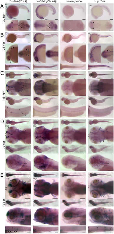
Fig. 1 Expression pattern of tubB4b[Chr5] and tubB4b[Chr14] mRNA during WT zebrafish embryo development. Whole mount in situ hybridization with tubB4b[Chr5]-specific antisense (1st row), tubB4b[Chr14]-specific antisense (2nd row), tubB4b[Chr5]-specific sense (3rd row) and myo7aa antisense (4th row) RNA probes was performed in zebrafish larva from 20 hpf (A), 24 hpf (B), 48 hpf (C), 72 hpf (D) and 5 dpf (E). A: lateral view of whole larva (upper panel); dorsal view centered on otic vesicle (left bottom panel) and on eyes (right bottom panel). B: lateral view of whole larva (upper panel); lateral view of head (left central panel); dorsal view centered on otic vesicle (right central panel); dorsal view of tail (bottom panel). C: dorsal view of whole larva and head (2 upper panels); lateral view of whole larva and tail (2 lower panels). D: dorsal view of whole larva and head (2 upper panels); lateral view of whole larva and head (2 lower panels). E: dorsal view of whole larva (upper panel); ventral view of head (2nd to the top); lateral view of whole larva, head and tail (3 lower panels). oc: optic cup; ov: otic vesicle; arrowhead: macular sensory hair cells; e: eye; p: pronephros; oe: olfactory epithelium; cmz: ciliary marginal zone; tb: taste buds; ∗: neuromast. Scale bar = 150 μm.
Reprinted from Developmental Biology, 517, Smaili, W., Pezet, C., Marlin, S., Ernest, S., R391 human dominant mutation does not affect TubB4b localization and sensory hair cells structure in zebrafish inner ear and lateral line, 301-316, Copyright (2024) with permission from Elsevier. Full text @ Dev. Biol.