IMAGE

Fig. 9

ID
ZDB-IMAGE-241127-58
Source
Figures for Lanoizelet et al., 2024
Image
Figure Caption

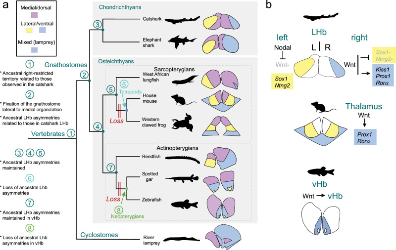
Fig. 9 Evolution of habenular asymmetries in jawed vertebrates.

a Evolution of the organization and the asymmetries of habenulae across vertebrates. Schemes on the right show the general subdomain organization of habenulae in members of the major vertebrate phyla. In gnathostomes, medial/dorsal territories are shown in purple, and lateral/ventral territories in yellow for territories of neuronal identity related to catshark Left-LHb or blue for territories of neuronal identity related to catshark Right-LHb. In the lamprey, territories related to the catshark medial and lateral right habenulae (respectively in purple and blue) are shown with the same color codes. Territories of mixed identity, co-expressing markers of medial and lateral right habenulae, are shown dotted in this species. Numbers at the nodes of the tree refer to the ancestral asymmetry profile inferred from comparative analyzes. The phylogenetic distribution of asymmetries suggests that a right restriction of neuronal identities related to those observed in the catshark lateral right habenula is an ancestral vertebrate feature (node 1 of the tree). The typical lateral to medial organization of habenulae, with lateral habenula asymmetries related to those observed in the catshark, were fixed in the gnathostome lineage, prior to its radiation (node 2). Lateral habenula asymmetries were maintained in chondrichthyans (node 3), as well as in ancestral osteichthyans, actinopterygians and sarcopterygians (nodes 4,5,7). They were independently lost in tetrapods (node 6) and neopterygians (node 8). b Differential deployment of an ancestral Wnt dependent regulatory module across jawed vertebrates. Functional analyzes in the mouse and in the catshark suggest that the Wnt signaling dependence of Prox1/Rorα expressions may reflect an ancient regulatory module, recruited to shape neuronal identities in the dorsal thalamus of tetrapods. In the catshark, the lateral right Kiss1 expression is also Wnt-dependent and the repression of Wnt by Nodal on the left side results in lateral Sox1 and Ntng2 expressions. In the zebrafish, Wnt signaling is required for ventral habenula formation, and possible later roles of the Wnt pathway in the elaboration of neuronal identities in this territory remain to be assessed. Abbreviations: LHb, lateral habenula; vHb, ventral habenula; L, left; R, right.

Acknowledgments
This image is the copyrighted work of the attributed author or publisher, and ZFIN has permission only to display this image to its users. Additional permissions should be obtained from the applicable author or publisher of the image. Full text @ Nat. Commun.