Figure Caption
Fig. 1
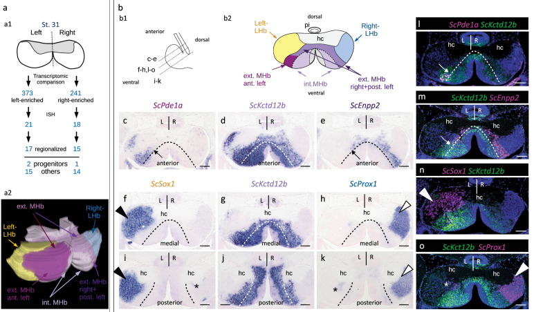
Developing catshark habenulae harbor major asymmetries both in lateral and medial compartments.
a Schemes showing the experimental strategy for the characterization of molecular asymmetries in catshark habenulae (a1) and the resulting 3D organization at stage 31 (a2). Anterior is to the left, dorsal to the top. b Schemes showing a left lateral view of catshark stage 31 habenulae, with section planes and levels indicated by dotted lines (b1), and the subdomain organization observed on a transverse section at a medial level (b2). c–o Transverse sections (dorsal to the top of each panel) after in situ hybridization (ISH) with probes for ScPde1a (c), ScKctd12b (d,g,j), ScEnpp2 (e), ScSox1 (f,i) and ScProx1 (h,k), and after fluorescent double ISH with probes for ScPde1a/ScKctd12b (l), ScKctd12b/ScEnpp2 (m), ScSox1/ScKctd12b (n) and ScKctd12b/ScProx1 (o). For fluorescent double ISH, signals for ScKctd12b are shown in green and those for ScPde1a, ScEnpp2, ScSox1 and ScProx1 in magenta. Sections (c–k) were obtained from the same embryo. Black and white arrowheads in (f,h,i,k) respectively point to major lateral territories of ScSox1 and ScProx1. White arrowheads point to major ScSox1 and ScProx1 major lateral territories in (n) and (o) respectively. Asterisks in (i,k,o) show contra-lateral minor posterior territories. Dotted lines in (c–m) delimit external and internal subdomains of the medial habenula, as inferred from the inner boundaries of ScPde1a and ScEnpp2 territories. Thin arrows in (c,e,l,m) point to the boundary between the complementary territories of ScPde1a and ScEnpp2 within the external MHb subdomain. Color code in (a2): yellow, Left-LHb; light purple, internal MHb; magenta, anterior, left-restricted component of external MHb; dark purple, right, plus posterior-left components of external MHb; blue, Right-LHb; hatched, pseudo-stratified neuroepithelium containing neural progenitors. Color code in (b2): same as in (a2); hatched, neural progenitors. The same ISH profiles were consistently obtained for each gene on at least five different specimens. Abbreviations: ant., anterior; post., posterior; ext., external; int., internal; MHb, medial habenula; LHb, lateral habenula; hc, habenular commissure; pi, pineal stalk; L, left; R, right; st., stage. Scale bar = 100 µm.
Acknowledgments
This image is the copyrighted work of the attributed author or publisher, and
ZFIN has permission only to display this image to its users.
Additional permissions should be obtained from the applicable author or publisher of the image.
Full text @ Nat. Commun.