
Image
|
Figure Caption
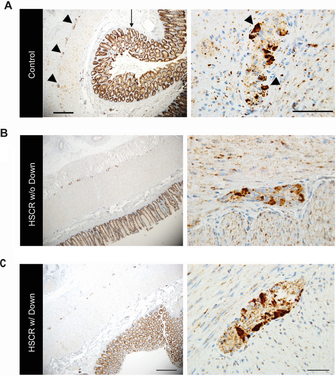
Fig. 2 ATP5PO is expressed in the myenteric plexus of the human colon. A. Intestine of a healthy individual showing ATP5PO staining in the myenteric plexus (arrowheads) and the epithelium (arrow). Scale bars represent 100 μm. B. Representative images of the (ganglionic) intestines of a HSCR patient without DS, and C. a HSCR patient with DS. Similar expression patterns of ATP5PO were detected. Scale bars represent 100 μm (left panels) and 50 μm (right panels); n = 3 per group.
Acknowledgments
This image is the copyrighted work of the attributed author or publisher, and
ZFIN has permission only to display this image to its users.
Additional permissions should be obtained from the applicable author or publisher of the image.
Reprinted from Biochimica et biophysica acta. Molecular basis of disease, 1870(3), Kuil, L.E., Chauhan, R.K., de Graaf, B.M., Cheng, W.W., Kakiailatu, N.J.M., Lasabuda, R., Verhaeghe, C., Windster, J.D., Schriemer, D., Azmani, Z., Brooks, A.S., Edie, S., Reeves, R.H., Eggen, B.J.L., Shepherd, I.T., Burns, A.J., Hofstra, R.M.W., Melotte, V., Brosens, E., Alves, M.M., ATP5PO levels regulate enteric nervous system development in zebrafish, linking Hirschsprung disease to Down Syndrome, 166991, Copyright (2023) with permission from Elsevier. Full text @ BBA Molecular Basis of Disease