
|
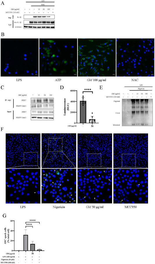
Fig. 3 GM suppresses the ATPase activity of NLRP3. (A) LPS-primed J774A.1 cells were treated with GM for 2 h and activated with imiquimod (200 μM) for 1 h. Supernatants and lysates were assessed via immunoblotting. (B) LPS-primed J774A.1 cells were treated with GM or NAC and activated with ATP (5 mM) for 10 min. ROS levels were fluorescently detected with DCF-DA solution (20 μM). (C) NLRP3-myc vector-transfected 293FT cells were treated with GM for 2 h. NEK7-NLRP3 interactions were confirmed using co-immunoprecipitation. (D) NLRP3's ATPase activity under GM treatment was determined via luminescence using an ADP-Glo assay. (E) Pellets of THP-1 cells were cross-linked using DSS (25 mM) and detected via immunoblotting. (F) LPS-primed J774A.1 cells were treated with GM for 2 h and activated with nigericin (10 μM) for 30 min. Representative immunofluorescence images of ASC speck formation were confirmed using a confocal laser-scanning microscope (Carl Zeiss, LSM710). (G) Graph showing the counts of ASC specks.