
|
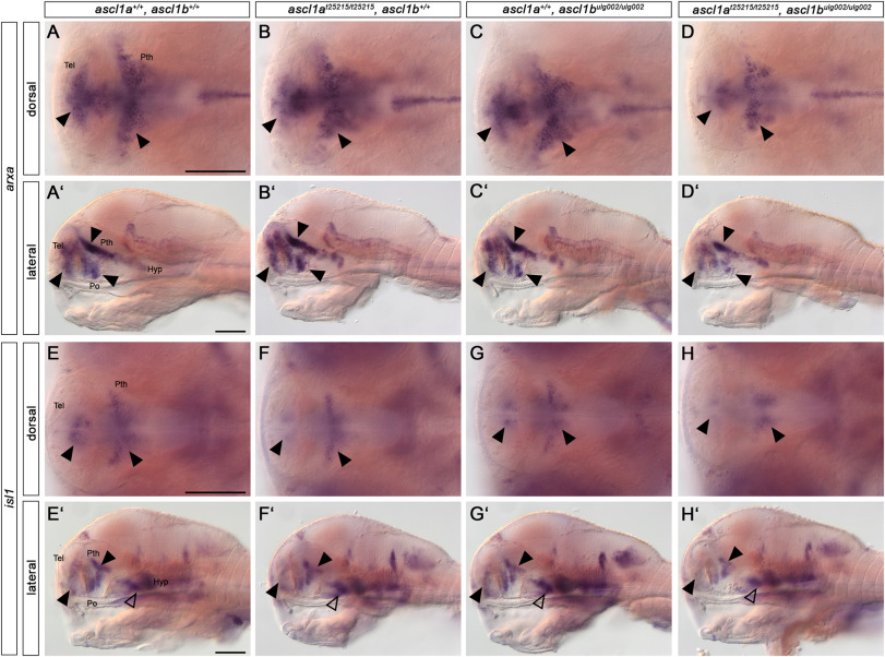
Fig. 7 Ascl1 regulates downstream effectors during dopaminergic differentiation. (A-H′) Whole-mount in situ hybridization for arxa (A-D′) and isl1 (E− H′) expression in 96 hpf embryos. Dorsal (A–H) and lateral views (A′-H′) of wildtype, single and double ascl1a and ascl1b mutant embryos. Anterior is to the left. Loss of Ascl1 function leads to a reduction of arxa expression (A-D′) in the telencephalon, prethalamus and preoptic region (arrowheads). The expression of isl1 (E-H′) is reduced in the telencephalon, prethalamus and hypothalamus (arrowheads) in double mutants compared to wildtype and single mutants. For numbers of analyzed embryos see Supplemental Table S2. Abbreviations: Hyp, hypothalamus; Po, preoptic region; Pth, prethalamus; Tel, telencephalon. Scale bars: 100 μm.
Reprinted from Developmental Biology, 505, Altbürger, C., Rath, M., Wehrle, J., Driever, W., The proneural factors Ascl1a and Ascl1b contribute to the terminal differentiation of dopaminergic GABAergic dual transmitter neurons in zebrafish, 587458-74, Copyright (2023) with permission from Elsevier. Full text @ Dev. Biol.