IMAGE

Fig. 6.

ID
ZDB-IMAGE-240224-12
Source
Figures for Dong et al., 2024
Image
Figure Caption

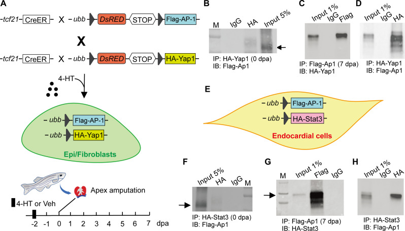
Fig. 6. AP-1 interacts with Yap1 and Stat3 in Epi/FBs and ECs, respectively.

(A) Schematic of 4-HT–induced Epi/FB-specific expression of tagged AP-1 and Yap1. The hearts were harvested at 0 and 7 dpa for Co-IP, respectively. (B) Co-IP using uninjured ventricles of the triple transgenic line TgBAC(tcf21:Cre-ERT2); Tg(ubb:loxp-dsRed-loxp-Flag-AP-1); Tg(ubb:loxp-dsRed-loxp-HA-Yap1). Yap1 was immunoprecipitated with anti-HA antibody, and AP-1 was detected by anti-FLAG antibody. A control IgG was used as the negative control for immunoprecipitation. (C and D) Reciprocal Co-IP of AP-1 and Yap1 in Epi/FBs at 7 dpa. IgG was used as the negative control. (E) Schematic of 4-HT–induced EC-specific expression of tagged AP-1 and Stat3. (F) Co-IP of AP-1 and Stat3 in ECs under uninjured conditions. (G and H) Reciprocal Co-IP of AP-1 and Stat3 in ECs during heart regeneration. AP-1 was immunoprecipitated/detected by anti-FLAG antibody, and Stat3 was detected/immunoprecipitated with anti-HA antibody. Control IgG was used as the negative control for immunoprecipitation.

Acknowledgments
This image is the copyrighted work of the attributed author or publisher, and ZFIN has permission only to display this image to its users. Additional permissions should be obtained from the applicable author or publisher of the image. Full text @ Sci Adv